Forums › Nd:YAG lasers › General Nd:YAG Forum › Removal of Twin Soft Tissue Lesions
- This topic is empty.
-
AuthorPosts
-
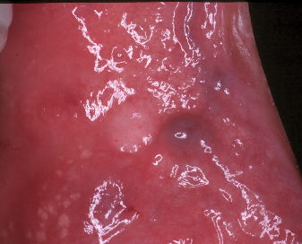
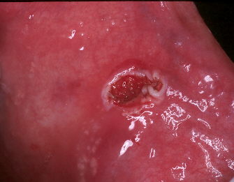
BenchwmerSpectatorA 65-year old male presents with two soft tissue lesions on buccal mucosa opposite right molars.
Diagnosis: Hemangioma and Irritation Fibroma
Treatment will consist of Ablation of hemangioma and excisional biopsy of fibroma
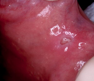
Free running, pulsed Nd:YAG laser (PerioLase) using 360 micron contact fiberFirst: Hemangioma will be entered with contact fiber, settings 3.0W 20Hz 150usec, dark blood is released, partial ablation of the vessel is done to prevent blood from repooling, and to allow healing. See small round entry wound. Less than 10 seconds exposure duration.

Then Fibroma is completely excised 3.0W 50Hz 150usec, placed in biopsy bottle. 20 seconds exposure duration.
No bleeding.
Placed on saline rinses.
Post Op photo taken at one week. Good healing of fibroma site taking place, resolution of hemangioma. Patient needed no pain meds.
Will re evaluate healing at 3 month recall.
AnonymousGuestJeff, how large was the fibroma? can I assume lots of air cooling since there is no brown tissue margin like you sometimes see w/ ndYAG?
Nice case.
BenchwmerSpectatorThe fibroma was under 5mm in width.
I place tension on the fibroma by pulling using a high quality, very thin tipped, tissue plier.
Start on one edge, the 50Hz setting increases the cutting speed. pull the fibroma as you cut.
Hi speed suction to remove the plume, no water or air.
Keep the tip moving,across the fibroma margin as it pulls away.
Be quick. Then no charring.
The Nd:YAG has no bleeding during or afterwards, Using the Erbium wavelength I have to then deal with an open, bleeding wound or alot of charring.
The Nd:YAG was the instrument of choice for this case.
Jeff
Robert Gregg DDSSpectatorJeff,
Nice case.
You have all the elements:
1. Tension
2. Edge and undermine
3. Quick
4. High Hertz (hot glass)
5. Lift lesion and separate
6. Dipfiber in and out to allow tip to reheat after quenching in tissue.Bob
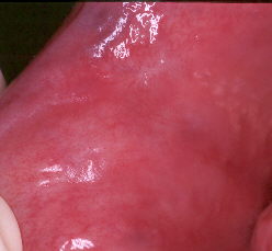
BenchwmerSpectatorFollowing is a 3-month post-op photo of the buccal mucosa after Hemangioma and Fibroma removal.

Complete healing, no re-occurence of either lesion.
Jeff
Glenn van AsSpectatorThe forum was down but sheesh ……great cases when people return to it.
Neat stuff and nice to see how the Nd Yag does on these.
Interesting email from one of my friends Sonny who works for Deka and is interested in my insights for a while on their lasers. I would imagine that this would involve their superpulsed CO2 and or their erbium.
I am interested in this kind of stuff as every experience teaches me the value of alternative wavelengths. There really isnt a one wavelength does it all mentality here and Jeff you have nicely showed how the NdYag can be very effective for hemangiomas and also for fibroma removal.
Great stuff there.
Glenn
-
AuthorPosts
