Forums › Nd:YAG lasers › General Nd:YAG Forum › Implant
- This topic is empty.
-
AuthorPosts
-
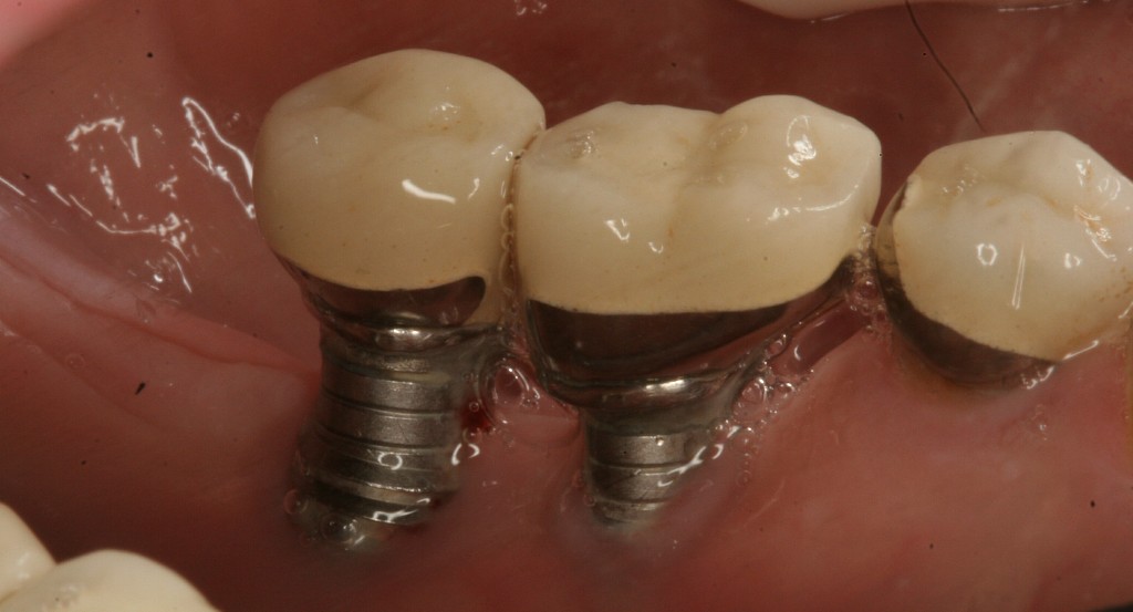
dkimmelSpectatorSo what do you think? LANAP stand a chance on this one?
[img]https://www.laserdentistryforum.com/attachments/upload/implantlanap1.JPG[/img]
doctorbruSpectatorDavid,
I have only done a few LANAPs so far since my september bootcamp.When we were at bootcamp and tough questions like this came up we heard ” These are the kind of questions for Day 4 and Day 5″. Since you have already completed those, Bob and Del may have to have a Day 6.I would love to here from someone( like Bob) who has used the LANAP to successfully resurrect dead implants. For that matter I would love to hear from anyone in this forum (too quiet in here- it’s spookey). With the right patient I could see myself attempting to save these implants. I would expect some sparks to fly close to that titanium..oughta be quite a show.
Bruce
Glenn van AsSpectatorThe crowns were never seated properly on the implants in my opinion (see the gap on the radiographs).
At least that is my opinion……now it could be the impression abutments werent seated or the final abutments not seated. There will be tissue in there now.
What kind of implants are these (corevent is my guess) and do they have an HA coating.
An erbium laser will absorb the HA and leave you with a titanium surface . It will also sterilize the area.
I have one case going on right now like this……waiting to see how it heals.
In my case the implant turns but the basket must have some bone in it as it wont pull up out of the way. I put the healing cap in and closed flapped it.
I will open flap it if it still turns in 3 – 4 months.
Its all experimental but in my case its on my mom who has the failing implant and I think she wont sue me!!
cya
Glenn
whitertthSpectatorGlenn,
I totally agree, abutments are definitely not seated and nothing will work if this isnt taken care of first….implants must be seated..
dkimmelSpectatorLooks like I need to do a Paul Harvey.
These are CoreVents that where placed in 1980. You are correct in that they are not fully seated. Some local wonder did the restorative work. Since they are Corevents from 1980–the abutments are cemented in place.. Very little chance of getting them out at this stage. Everything is rather high and dry at his point and even with LANAP this is a rather questional prognosis. She is 56 and just had a heart attack in Jan. Very nice school teacher with limited $$$. I am afriad that the implants are the least of her dental problems.We have a bridge on the lower left that is about to fall off and another bridge on the upper right that is placed on half of the tooth because the rest was sub crestal…..
My thoughts are to stablize the lower right for as long as possible until we can get her undercontrol…
Dentistry in the real world is so much fun…
doctorbruSpectatorDavid,
Thanks for posting this case.
What are your thoughts on how you plan to stabilize her ? Are you going to try the LANAP ? If so, what differences will you be thinking about using the protocol on implant vs natural teeth ? Will you be probing around the implant, firing the laser near the surface-what effect on the titanium would you expect?Can you clean the titanium? Would you use the piezo or hand scalers on the titanium surface ?
Sorry David for so many questions. I have several patients with similar problems and would like to know if I will be able to help them. I have zero experience with saving failing implants- usually refer them to the oral surgeon.
Bruce
AnonymousGuestThought you guys might find this useful-
J Oral Maxillofac Surg. 2005 Oct;63(10):1522-7. Related Articles, LinksSurface properties of endosseous dental implants after NdYAG and CO2 laser treatment at various energies.
Park CY, Kim SG, Kim MD, Eom TG, Yoon JH, Ahn SG.
Department of Oral and Maxillofacial Surgery, Oral Biology Research Institute, College of Dentistry, Chosun University, 421 SeoSeogDong, GwangJu City 501-825, Korea.
OBJECTIVES: Dental lasers have been used for uncovering submerged implants as well as decontaminating implant surfaces when treating peri-implantitis. The objective of this study was to compare the possible alterations of the smooth surface and resorbable blast material (RBM) surface implants after using NdYAG and CO(2) lasers at various energies. MATERIALS AND METHODS: Ten smooth surface implants and 10 RBM surface implants were used. Two smooth surface implants and 2 RBM surface implants served as a control group that was not lased. The remaining implants were treated using NdYAG and CO(2) lasers. The surface of each implant was treated for 10 seconds on the second and third threads. The smooth surface implants (group 1) were treated using a pulsed contact NdYAG laser at power settings of 1, 2, 3.5, and 5 W, which are commonly used for soft tissue surgery; the corresponding energy and frequency were 50 mJ and 20 Hz, 100 mJ and 20 Hz, 350 mJ and 10 Hz, and 250 mJ and 20 Hz, respectively. The group 2 RBM implants were treated using a pulsed contact NdYAG laser. The group 3 smooth surface implants were treated using a pulsed wave non-contact CO(2) laser at 1, 2, 3.5, and 5 W, and the group 4 RBM implants were treated using a pulsed wave non-contact CO(2) laser. Data were analyzed using scanning electron microscopy. RESULTS: The control surface was very regular and smooth. After NdYAG laser treatment, the implant surface showed alterations of all the surfaces. The amount of damage was proportional to the power. A remarkable finding was the similarity of the lased areas on the smooth and RBM surfaces. CO(2) laser at power settings of 1.0 or 2.0 W did not alter the implant surface, regardless of implant type. At settings of 3.5 and 5 W, there was destruction of the micromachined groove and gas formation. CONCLUSION: This study supports that CO(2) laser treatment appears more useful than NdYAG laser treatment and CO(2) laser does not damage titanium implant surface, which should be of value when uncovering submerged implants and treating peri-implantitis.
Note “contact”. Seems to indicate that, like endo, a distance from the target(4mm?) should be respected, as well as the angle. Bob, thoughts?
Robert Gregg DDSSpectatorRon,
Don’t know the dosimetry. How long were the implants exposed and in contact? Parallel or perpendicular?
CO2 is fine, nothing wrong with FRP Nd:YAG if used properly.
Bob
doctorbruSpectatorBob,
Is it correct to assume that a mostly end cutting fiber such as used on the FR:NdYag would be fine if kept parallel to the implant surface ?
Would it be prudent to wipe the tip more frequently to prevent any nonendcutting heat from building up on the fiber tip ?
Bruce
AnonymousGuestQUOTEQuote: from Robert Gregg DDS on 1:00 pm on Oct. 14, 2005
Ron,Don’t know the dosimetry. How long were the implants exposed and in contact? Parallel or perpendicular?
CO2 is fine, nothing wrong with FRP Nd:YAG if used properly.
Bob
Medline is still working on the article so I dropped one of the authors an email with the methods questions.
Glenn van AsSpectatorOne of the things to consider is that if you flapped the case that the surface of exposed hydroxyapatite could be easily removed with the erbium lasers. THe hydroxyapatite is readily absorbing the erbium lasers and it will do little to the titanium surface. Water spray keeps everything cool so it might be the way to go in my opinion as the right wavelength for this type of failing implant. The wavelength will strip of the HA and will sterilize and not heat up bone.
The ongoing problem of course is the gap between superstructure and the implant itself.
I think that you will see with time that erbium lasers will become a treatment of choice for failing implants. Lots of research looking at this now.
Glenn
dkimmelSpectatorAs odd as it may seem I am not to concerned about the fit or lack of fit at the implant post interface. It is alomost high and dry at this point and I will probably just seal it as best possible. I don’t even want to think about trying to remove this cemented abutment….
I am more concerned about her occlusion as the doc paid as much attention to the occlusion as he did in seating the crowns and the abutments…..
That said, I still have a wonderful defect..
Several concerns come to mind.
The biggest is removing the tissue that is in the basket.Pretty hard to remove this and keep the Nd:YAG away from the implant. This would be a non issue with an Er. However, this would mean I would have to open the site. Not really what I would want to do if I expect to get the full benifit of the Nd:YAG!!! RAther a CATCH 22.
Which brings me to the real question.
Do I really want to try and get the osseous defect to fill in???
These are not two implants that I would like to maintain for a very long time. As it is now they can be farely easly removed and block grafts placed. If I do get any resolution of the defect I have just made this a harder job…
Glenn van AsSpectatorDavid I will tell you that the distal implant will be a piece of cake, the mesial a real huge headache.
I just tried to remove one on my mom which was exactly the same Corevent HA coated implant. It spun but wouldnt come out.
I finally closed flapped around the implant with the erbium, removed the superstructure and then put a healing cap in.
Will see in 3months and most likely then will flap it to see what can be done. I could spin the implant but no way could I get it up…..
It looked just like your mesial one on the radiograph.
Glenn
Robert Gregg DDSSpectatorQUOTEQuote: from doctorbru on 2:20 pm on Oct. 14, 2005
Bob,Is it correct to assume that a mostly end cutting fiber such as used on the FR:NdYag would be fine if kept parallel to the implant surface ?
Would it be prudent to wipe the tip more frequently to prevent any nonendcutting heat from building up on the fiber tip ?
Bruce
Yes, it is end-firing, but th scatter in the tissue is circumferential around the tip, but making the intensity much less of course (a good thing in this instance).
No need or concern to remove the protein B/U. Just not hot enough.
Thanks Ron.
Bob
mkatzSpectatorto remove teh spinning corevent is easy – section it, just as you would a tooth. High speed carbide fissue burs with good coolant water spray. Bone removal not required.
-
AuthorPosts
