Forums › Laser Treatment Tips and Techniques › Soft Tissue Procedures › fibroma removal
- This topic is empty.
-
AuthorPosts
-
whitertthSpectatorroutine excisional biopsy of fibroma on left cheek…. .5 watts 14/8 emla topical….pretty good hemostasis as well…..enjoy
whitertthSpectatorhere are the photos sorry…..
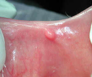

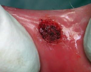
Glenn van AsSpectatorHI Ron: neat pics, did you do this with the erbium?
You got pretty good hemostasis with this one too.
All the best. ….nice job.
glenn
whitertthSpectatoryes dome with the waterlase at .5 watts 14/8 after the removal i went back at the same power without water and very little air to apply my “laser bandaid” coating the defect with laser energy…i pressed on void with gauze for 20- 30 seconds and thats what i got…
AnonymousGuestThought I’d add a fibroma case also.
5mm diameter
er,cr:YSGG 1.0 W EMLA 11/7Preop
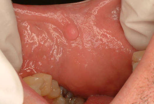
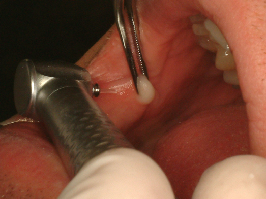
Postop

1 week

Ok, a few questions for you all.
Should I have been more invasive and gone deeper?
In this case after the piece was removed I went back and ‘resurfaced’Biopsy?
Pt could pin point cause from trauma.
Any suggestions?
ASISpectatorHi, Fellow Rons,
Very nice photos. Interesting that there’s no bleeding from the second case. Is that why you think it should be done a bit deeper?
Andrew
AnonymousGuestQUOTEQuote: from ASI on 11:44 am on May 19, 2003
Hi, Fellow Rons,Very nice photos. Interesting that there’s no bleeding from the second case. Is that why you think it should be done a bit deeper?
Andrew
Actually, most of the cases I’ve seen posted,here and elsewhere, seem to look like they are more cratered afterward. I didn’t create a crater with this one and was wondering if there is any long term diffrence.
I’ve never had any bleeding to contend with on fibroma removals, just once in awhile on frenectomies.
SwpmnSpectatorLooks real nice, Ron.
I don’t think any need to go deeper or biopsy.
Al
BenchwmerSpectatorIs this a WaterLase only topic?
If not stated differently I guess we are to assume WaterLase?
I though we were going to give out treatment parameters, tip size, duration, etc.
I never see Hertz posted in WaterLase posts, even if it is fixed, it would be helpful in treatment interpretation.
Ron,
What size tip did you use?
I would place tissue in biopsy bottle, evaluate healing and even then keep to see if insurance will pay w/o biopsy report, never know when you need that tissue.Jeff
AnonymousGuestQUOTEQuote: from Benchwmer on 8:16 pm on May 19, 2003
Is this a WaterLase only topic? If not stated differently I guess we are to assume WaterLase?Only if no other wavelengths get posted 😉
Jeff, I always try to post wavelength rather than brand.QUOTEI thought we were going to give out treatment parameters, tip size, duration, etc. I never see Hertz posted in WaterLase posts, even if it is fixed, it would be helpful in treatment interpretation.Sorry, I left out G4 tip and the Waterlase (er,cr:YSGG )is fixed at 20H. Tx time approx. 1 minute. My fault for posting between patients.
Time for some other wavelength users to post similar cases!
BenchwmerSpectatorRon,
Here is how it is done with a pulsed, FR, Nd:YAG, contact fiber, 3.0W 20 Hz 110usec, less than a minute.
3 point infiltration, 2% Carbocaine w/ 1/20,000 Levordefrin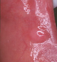
Before treatment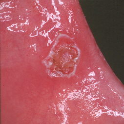
Immediately after treatment
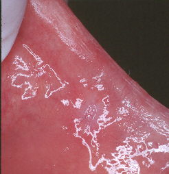
two weeks post-op
whitertthSpectatornice job!!
AnonymousInactiveI look at the pictures and know this is great fun learning all these new methods. This is such a tremendous forum to share on.
I have found that the difference in wavelength (and therefore the tissue interaction) of the laser made a significant difference in the removal of a fibroma as you have presented. With regards to the depth of the cut you make – the fibroma should be the determining factor. With the erbium or the Diode you must decide where you will make your cut. Many times the tissue will feel the same and it is hard to differentiate what you’re removing from tissue you wish to keep. I also see this dilemma in the questions that are posed. With the FR Nd:YAG used in a selective ablation mode these tissues are recognizable and are able to be distinguished one from the other and separated. Thus you are able to remove the complete fibroma without sacrificing any additional tissue unnecessarily. Now that Bob has the camera and scope set up we will post the next one of these we remove – I hope he lets me use the camera!
Robert Gregg DDSSpectatorNOPE!! uh, ah. ain’t sharin’….:biggrin:
BenchwmerSpectatorHere is my first fibroma removal using the OpusDuoE.
10 Hz 350mJ using a 800micron tapered sapphire contact tip for less than 30 sec.
LA 2% Carbocaine w/ 1/20,000 Levordefren, tripod technique (3 drops in triangle surrounding lesion in mucosa)Before
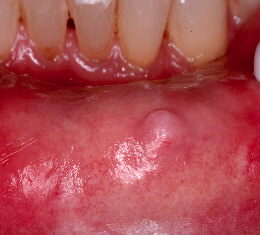
Immediately after

Next time I’ll use a 200 or 400micron tapered tip for more focused tissue response.
Jeff
-
AuthorPosts
