Forums › Laser Treatment Tips and Techniques › Soft Tissue Procedures › Laser cosmetic gingival contouring/crown lengthening
- This topic is empty.
-
AuthorPosts
-
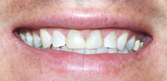
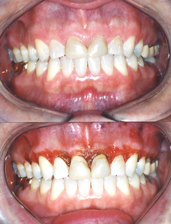
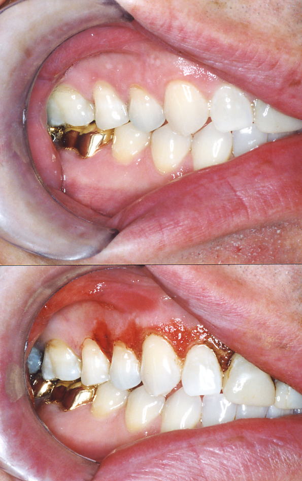
RodSpectatorThis is a case I did this morning. I posted it because the patient is a dentist and recognizable Townie. I’ll ask him if he minds me giving his name.
Please note that he ALREADY had a periodontist do cosmetic contouring/crown lengthening, and the first photo of his smile was taken this morning before we started (this is AFTER the periodontist’s handywork). The periodontist told him that this is the best he could do without flapping and osseous.

You can see this was not the case. This was done by laser. A new sulcus was created also with the Biolase LaserSmile diode laser. I used a thin small instrument to create the sulcus about 1.5-2 mm depth, and then used the laser at 1watt continuous, dragging through the ‘new’ sulcus of each tooth. (this will inhibit re-growth of the tissue)
You can imagine that if I only ‘contoured’ up the gingival margin and left it that way, we’d have a blunt, THICK marginal gingiva. Therefore note that after the contouring, the gingiva was ‘thinned/festooned’.


You don’t see a post-op of his smile because he was numb after the procedure and his ‘smile’ looked stupid 😉
Think this looks painful? Think again. This case was desensitized (similar to what we’d do to treat a canker sore) with the laser. Here’s an email I just got from this Townie patient:
“you wanted to know so you can tell patiens so heres the scoop as of ..well its 1:30 and ZERO PAIN and numbness totally worn off,,,
rite when i left office i DID HAVE some very mild discomfort for about an hour but that passed …i did place rembrandt junk on gums too which helped earlier…but rite now i am touching gums and zero pain!!!! and my wife couldnt belive how good an dlonger my teeth look
great job doc and enjoy your weekend”
(Edited by Rod at 3:31 am on Mar. 22, 2003)
(Edited by Rod at 3:34 am on Mar. 22, 2003)
Glenn van AsSpectatorNeat stuff Rod and I for one really enjoy seeing you post this case.
The more the merrier.
Please show us the healing photos……whether you get rebound or if it stays the same.
Great stuff for the patient.
Glenn
One suggestion if you want dentists to not be critical………
Take a photo with a probe in the tissue prior to doing the laser work after you have anesthetized and then they cant say you didnt sound the bone.
Glenn
RodSpectatorHey Glenn,
What’s “sounding the bone”? ;~) LOL!!!
Rod
SwpmnSpectatorRod:
I’ve already sent my comments on the case in the other forum. Thanks for posting here.
Al
PatricioSpectatorRod,
Could you say a little more on how you created the new sulcus with the thin instrument? Did you remove bone with your laser? Will you anticipate some recession from the festooning as the tissues heal?Pat
RodSpectatorQUOTEQuote: from Patricio on 7:56 pm on Mar. 25, 2003
Rod,
Could you say a little more on how you created the new sulcus with the thin instrument? Did you remove bone with your laser? Will you anticipate some recession from the festooning as the tissues heal?Pat
Hi Pat,
We know that the ‘biologic width includes some fibrous AND epithelial attachment. Once you’ve removed soft tissue down to the fibrous attachment, the epithelial attachment is all gone. The fibrous attachment is pretty tenacious stuff, and left as-is will tend to promote some re-growth of tissue, which we do NOT want.
So what I do is take an explorer or perio probe and run it down, back and forth, between the fibrous gingival tissue and the root. Then I take the tip of my Biolase LaserSmile/Twilight laser into this ‘sulcus’ that I just opened up and run it back ‘n forth too. This will remove the fibers so that we have a better chance of forming an epithelial attachment along with re-establishing some fibrous attachment (therefore a new ‘biologic width’ with both fibrous and epithelial attachment). This means that the tissue doesn’t have to try to re-grow height to create an epithelial attachment.
No, in this case I did not need to remove bone, based on the depth of the bone sounding. By the way….”bone sounding”….I’ve always wondered what bone REALLY ‘sounds’ like. 😉
And no, I do NOT anticipate ANY recession from the festooning. Actually I expect regrowth of the epithelial layer over the festooned fibrous gingival tissue — but not enough to make any difference. It will NOT recede because the remaining tissue is VERY fibrous (read ‘durable’).
Rod
Robert Gregg DDSSpectatorHi Rod,
This ought to be a very nice end result.:)
Would you explain your technique for festooning the tissues?
What laser device, fiber, “mode” did you use (activated versus non-activated fiber) and motion of the fiber did you use?
I’m asking because what you did is not as easy as it might appear, and a good discussion might be helpful to all.
I always get a little nervous using near-infrared lasers (diodes or pulsed Nd:YAGs) for thinning fibrous tissue on the facial since these wavelengths are poorly absorbed in fibrous and conective tissue.
I remember 4 years into using lasers–and thinking I was pretty experienced–(read that as cocky) and I treated a case like this. I used water to “cool” the tissue as I was “debulking” the fibrous tissue. Well, all I was really doing was cooling the tip of my fiber, and warming (read that necrosing) the fibrous tissue beneath, instead of removing it.:o
For two weeks (and it took about 48 hours to start hurting) my patient had a very painful recovery as the tissue died, sloughed off, and regrew!

The good news is that all the tissue regenerated, and it is now “bullet proof” tissue, in that the collagen that repaired in the area is very prolific and redundant tissue, making it “tense” not thickened or hypertrophied.
Anyway, this can be achieved with a very satisfactory and confortable outcome if one does not do what I did 8 years ago. And the tissue appearance looks as though Rod did it right!
By the way–I have a video tape of what I was doing on this patient/case–if anyone whats to see how NOT to do it! You can imagine I don’t show it very often. In fact, it has been gathering a lot of dust!! I guess it’s remarkable because it doesn’t look like anything harmfull is taking place……
Thanks for posting Rod.
Bob
RodSpectatorHi Bob,
Can’t remember if I went into the technique here, on the DentalTown message boards, or on the DentalTown case presentation site — guess it’s age-related brain fade, huh?
Anyway, I didn’t use a laser at all to festoon the tissue. Yes, the gingival margin area I used a diode with an itiated tip. But then to blend it in and do the festooning of the surface I used a wheel diamond in a highspeed. I use the flat end of the wheel, and tilt it just a little as I ‘paint away’ the tissue.
Then after the festooning (thinning and blending it into the new gingival margin) of the tissue, I used the Waterlase in exactly the same way I’d use it when treating a canker sore. I simply coated the surface a couple times at a low wattage and no water (such that the surface became white).
And just like a canker sore, it worked like a charm. The patient (who is a dentist himself, and a recognized Townie) has done great and emails me almost every day.
After a day he was a little alarmed that the papillas seemed to be rapidly re-growing. He had always HATED his thick, fat, bulbous papillas, and he was totally flipped out at the thin, knife edge papillas we ened up with.
So when he saw the tissue growing rapidly after a day, he freaked. I told him not to worry. Hypertrophy doesn’t happen that fast. I told him is was simply a bit of edema and would go back to where we’d put it (although I’d taken it away more than I wanted it, in anticipation of growing a new epithelial covering, which it will do).
Anyway, after about three days the edema did go away and the tissue went right back.
But as far as pain, it’s not been an issue.
I haven’t seen him since the surgery, but he is totally excited about the outcome. I should be seeing him probably within the next week. I’ll take photos of the 2-3 week post-op and post them.
And yes, I’d agree. I’d never want to do that sort of festooning with a laser. The diamond is great because I can ‘feel’ what I’m doing and can sculpt VERY quickly. The festooning took me no more than two minutes, and probably less.
But without the laser to desensitize those cut nerve ends after the festooning, holy moly — PAIN would have been the result — BIG TIME!!
By the way, the post-op photo was taken AFTER desensitizing the surface with the Waterlase. Even though the Waterlase turns the surface white as you use it, as soon as it gets wet, the white disappears.
Rod
(Edited by Rod at 1:31 am on April 3, 2003)
Andrew SatlinSpectatorRod,
Laser dentistry is very new to me. I do however have alot of experience in periodontal surgery. Crown lenghtening or any preprosthetic surgery in the esthetic areas are particularly challenging. I am still unclear about your technique and your rationale. It seems that you : 1- performed 2-3 mm of laser gingivectomy
2- used a probe / explorer for blunt dissection into the connective tissue attachment
3-ablated the connective tissue with the laser
4-gingivoplasty with diamond burs
How does this create a new sulcus? could you have accomplished the same procedure with a 15 blade and a diamond bur?
Aren’t you just violating the biologic width and alowwing the periodontium to heal on its own?
I am not an expert on wound healing but it seems that if you did not leave enough space for epithelium, connective tissue and sulcus depth then the periodontium will reestablish its natural biologic width via attachment loss.
Cases like this one with thick tissue may be very forgiving. Others may not be.
I do not mean to sound critical. I am always looking for better ways to preserve papillae and obtain adequate crown lenghth.Andy
RodSpectatorHi Andy,
Some very good questions. Many of these have already been answered, but may have been answered on the DentalTown forum or case presentation area.
I’ll ‘quote’ you and then answer each quote below.
QUOTEQuote: from andy on 5:37 pm on April 12, 2003
RodQUOTELaser dentistry is very new to me. I do however have alot of experience in periodontal surgery. Crown lenghtening or any preprosthetic surgery in the esthetic areas are particularly challenging.Yes indeed they are — VERY. It’s easy to do more, but if you get too aggressive the first round, it’s more difficult to ‘put it back’.
QUOTEI am still unclear about your technique and your rationale. It seems that you : 1- performed 2-3 mm of laser gingivectomy
2- used a probe / explorer for blunt dissection into the connective tissue attachment
3-ablated the connective tissue with the laser
4-gingivoplasty with diamond bursFor the most part, yes, but actually #1 and #3 above seem to be saying the same thing — removal of ‘soft’ tissue with the laser.
QUOTEHow does this create a new sulcus?Actually, saying this creates a new sulcus is not exactly correct. What this does is help create epithelial attachment. Above the level of the bone we’re gonna have connective tissue attachment via fibers, epithelial attachment and sulcus. Sometimes the connective tissue fibrous attachment is wider than others. If you start out with a very wide connective tissue attachment, you can usually reduce this width of attachment successfully.
You will commonly find this in cases of ‘gummy’ smiles. In cases like this, often the bone is where it would be even if this patient didn’t have a gummy smile. Whereas the ‘average’ patient would have a narrower band of fibrous tissue, the gummy smile patient may have a much wider band. In a case like this you can reduce the width of the band of firm fibrous attachment without the amount of rebound that you’d expect if you encroached on the bone level.
One key is getting rid of some of those fibers. By severing the fibers, and then use of the laser, you’re more likely to establish some epithelial attachment — whereas if you left that tissue attached via fibers, you’d be more apt to grow more tissue (rebound) to establish the epithelial attachment portion of the ‘biological width’.
QUOTEcould you have accomplished the same procedure with a 15 blade and a diamond bur?Yes, absolutely, and I have many times. But there are two problems with that. One is pain. The post-op pain is quite significant without the laser. The other is that the fibers (which we intentionally sever) are more likely to re-establish without use of the laser. But yes, I’ve done this numerous times with instruments other than the laser.
QUOTEAren’t you just violating the biologic width and alowwing the periodontium to heal on its own?I’m very confused about this question, and unsure of what you’re getting at. I’m certainly taking away some of the biologic width, it that’s what you mean. However ‘violating’ is a different thing. I’d say that ‘violating’ the biologic width would be removing more than you need to have a healthy biologic width. I wouldn’t call removal of ‘excess’ biologic with ‘violating’. Is that what you meant? True violation of biologic width simply doesn’t work — whether or not you use a laser.
However, if the patient starts out with excess biologic width, you can successfully reduce this with and without a laser, although it’s easier with a laser. The main thing is to retain a biologic width that is acceptable.
Before I forget, take a look at the DentalTown case presentation at another case I posted in response to similar questions. It’s a case where I removed a large amount of biologic width over one of the centrals. We could have been more ‘perfect’ by doing a touch of osseous in a couple areas, but this is presented to show a 9 month post-op.
QUOTEI am not an expert on wound healing but it seems that if you did not leave enough space for epithelium, connective tissue and sulcus depth then the periodontium will reestablish its natural biologic width via attachment loss.Huh? Again not sure what you mean by ‘attachment loss’. Are you saying that the biologic width will reestablish itself via bone resorption? In other words, are you saying that if you violate the minimum biologic width, and place restorations to the level of the new gingival margin, the gums will not grow back up, and instead will reestablish via bone resorption? This may very well be the consequence, however there would be a chronic inflammation during this period, which may be more extended in some patients than others. I have found that very slight violations of minimum biologic width are usually tolerated if they occur only here and there, but not if significant areas are involved.
QUOTECases like this one with thick tissue may be very forgiving. Others may not be.Again, not at all sure of what you’re getting at. Are you saying that cases with thick tissue are forgiving, but cases without thick tissue are NOT forgiving? If this is what you’re saying, you’re entirely correct. That’s why I’ve mentioned that we first established where the bone was before deciding what technique to utilize. Given that we started out with an excessive biologic width, to use your phrase, we understood that the case would be ‘forgiving’ and not require osseous.
Or are you saying that some cases of thick tissue will be forgiving, and some cases of thick tissue will NOT be forgiving?
Actually, there was a little more behind our decision making process than meets the eye, and it may turn out that we’ll regret not doing some osseous now. In the beginning, we anticipated doing the cosmetic work on the gums, and then a combination of porcelain jackets and veneers all around. We were to gain some tooth length via reduction of the gingiva, as well as lengthening of the incisal edges in porcelain. Given this scenario, the amount of gingival contouring would have been perfect. HOWEVER, the patient (who is himself a dentist) has now changed his mind and decided to ONLY treat the two centrals. Because of this, lengthening the teeth will not be an option, and even more gingival removal would have been ideal.
As mentioned before, we ALWAYS anticipate growth of some gingiva, and therefore when we do the procedure, we must ‘overdo’ it a bit. We remove epithelial tissue down to the connective tissue. We WILL ALWAYS have regrowth of epithelial tissue over that rather durable connective tissue. So we deal with a ‘controlled’ amount, or ‘anticipated’ amount of rebound.
However, I’ve found that most often the laser will keep the anticipated rebound less than if the laser is not used.
For some reason several have misunderstood the purpose of posting this case — thinking that I am saying that by using a laser, we can successfully violate the minimum biologic width. I’m not sure why some are thinking this. It simply is not true. It is true that the laser will usually control rebound better than similar procedures without lasers — therefore making the procedure more predictable. But violating minimum biologic width? No.
The purpose was to show how such a case looks with a laser as the instrument, as well as to show that sometimes dentists go in and do osseous when they actually could have avoided it.
This patient was treated to remove excess gingiva by a periodontist within a few months before I saw the patient. The periodontist told the patient that to achieve more results, osseous would be necessary. Upon exam, given the apparent width of connective tissue attachment, I had a suspicion that we could have a better result without osseous. However, we were prepared to go either way, depending on how the bone sounding went after anesthetic was used.
I do not know who the periodontist was. I don’t know if he does much of this sort of stuff. I hate to say it, but what went through my mind is that this dentist (the patient) may refer to this periodontist. And because of this, the periodontist may have done this as a freebee for the referring dentist patient. And maybe he just didn’t feel like putting forth the effort to really take his time and go the distance. Maybe, maybe not. It’s not a discussion I really wanted to get into much with the patient.
QUOTEI do not mean to sound critical. I am always looking for better ways to preserve papillae and obtain adequate crown lenghth.OK, NOW you touched on something that is a ‘pet’ of mine — preservation of papillae. I don’t know if I mentioned it on this forum, but I think I did on one of the DentalTown forums. Cracks me up when I read an article where they show how to do cosmetic crown lengthening, and utilize the ‘papilla preservation’ technique. Boy!! You talk about ‘hit ‘n miss’!!
When I do flaps to do osseous, I NEVER use this papilla technique. I make my incision without going into the papilla. Yes, I ‘filet’ off the most facial portion of the papilla, but not even up to the tip, and certainly I don’t go into the interproximal space whatsoever. I do the osseous reduction and apically position the flap. Now I’ve ended up with the papillas extending farther from the gingival margin than when we started. The papilla tips has not changed, but the gingival margin has. In these cases, you never lose a papilla, but most often you need to later go in and do ‘touch-up’ contouring.
Most often you will do some facial osseous, but really don’t need any interproximal bone reduction — but sometimes you do. In cases like that, I do the same approach, but once the facial osseous reduction is finished, if I need to ‘blend it’ into the interproximal, I simply use the Waterlase and remove a little bone UNDER the papilla going into the interproximal. Works like a charm.
Anyway Andy, great questions. Believe me, I’m NOT ‘the authority’ on this. I’ve done cosmetic gingival contouring for about 23 years now — started many, many years before it was ever spoken of in the journals — way before periodontists started thinking about it as a legitimate reason for treatment. So I am somewhat experienced in it, but NOT any sort of ‘authority’ by a long shot. I too am always looking for better ways.
Thanks.
Rod
(Edited by Rod at 12:25 am on April 13, 2003)
Andrew SatlinSpectatorHi Rod
Thank you for your detailed reply. It seems we agree on many points. A couple of things still need to be clarified.
How do you determine removal of “excess” biologic width vs. violation of healthy biologic width. I have never actually heard the term “excess ‘ biologic width.Re: attachment loss –that is exactly what I was saying. If you violate the biologic width you may very likely get a chronic inflammatory response. I am sure you have seen it over the years.
Re: cases with thick tissue being forgiving. Again you understood my point perfectly. Thick periodontiums are very forgiving.
Re: Papillae preservation–We should probably talk on the phone about this one. I could not agree with you more. I acually use the same technique if I have to get access to bone. I have had minimal success with the “palatal curtains” and the old published methods. Actually, if you look carefully at those newer “cosmetic” crown length articles they are very misleading. I often reccommend orthodontic extrusion in isolated cases. Have you tried that?
Thanks again Rod –talk to you later
Andy
RodSpectatorHi Andy,
Very glad to see we think alike. As far as having see the chronic inflammatory response from violation of the biologic width — absolutely, I’ve seen it — and sometimes by my own hand (well, I guess that’s how we must learn sometimes, huh?)
As far as the term ‘excess biologic width’, hey, I made up that term myself — pretty cool, huh? LOL!! Actually, these are simply the cases you mentioned that have the excessive thick tissue — ‘thick’ meaning a wider band of fibrous tissue. If you notice, they’re forgiving, but ONLY forgiving if you do not invade that minimum width of biologic width. Given that, in my mind, they start out with ‘excessive biologic width’, I personally think this is exactly why they’re forgiving.
Andy, I see you’re in So. Cal. So am I. Email me and lemme know where you are. My email is [email=”drrodger@cox.net.”]drrodger@cox.net.[/email]
Rod
-
AuthorPosts
