Forums › Laser Treatment Tips and Techniques › Soft Tissue Procedures › Stable Fibrin Clot Immediate & 7 Day Post Op
- This topic is empty.
-
AuthorPosts
-
Robert GreggParticipantHi Everyone,
Here’s a “Laser Periodontal Therapy” case I started last week.
This photo series demostrates the key element in treating ALL types of periodontal disease case types in only ONE treatment visit. That is, as opposed to treating a pocket or quadrant in a series of sequential visits, one week apart.
They KEY is getting a stable fibrin clot to form immediately post-op, and before the patient is dismissed.
Profound anesthesia is ALWAYS used for intial treatment.
[img]https://www.laserdentistryforum.com/attachments/upload/MarkXray.JPG[/img]
Pre-Op X-ray. Can a pocket be a VERY thick PDL space?
[img]https://www.laserdentistryforum.com/attachments/upload/Markpoach1.JPG[/img]
4.00 watts, 20 Hz, 200 mj/p, 360 micron fiber, 650 usec PD @ 110 Joules per tooth
[img]https://www.laserdentistryforum.com/attachments/upload/Markpoach2B.JPG[/img]
Obtaining a predictable, controllable, reproducible and STABLE fibrin clot has been difficult for all laser devices (especially CW diodes with or w/o “activated” hot-glass effect), until we found the value of varying the pulse duration on a pulsed Nd:YAG. Extending the thermal “injury” time for just a few hundreds of microseconds made all the difference—not too cold, too short thermal exposure time, or too photo-disruptive, AND not too hot as to vaporize, cauterize, burn the blood proteins away (they are VERY thermally sensitive;-), OR sear or DRY out the tissue edges that would prevent “clotting closure” of the wound.
[img]https://www.laserdentistryforum.com/attachments/upload/Mark7day3.JPG[/img]
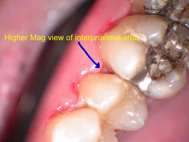
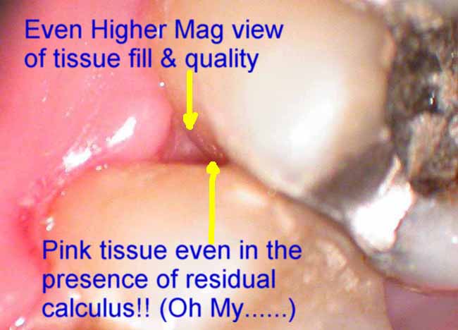
7 Day post-Op reveals fibrin clot persisting in the area still struggling to heal in the bicuspid region. The area that is healing the best, is the area which had the deepest pockets, most root surface areas involved and most negative tissue architecture. But it is important to note that the area still needing the fibrin coverage, is still showing the fibrin along with residual tissue redness and inflammation. So is that SLOW healing? Or something else???

Microscope shows higher magnification of the desirable fibrin and its appearance. No, it is NOT necrotic tissue. It also shows a better idea of how well the inner interproximal tissues are healed……sealed down below.
[img]https://www.laserdentistryforum.com/attachments/upload/Mark7dayHiResV4.JPG[/img]
Self explanatory

Self explanatory
[img]https://www.laserdentistryforum.com/attachments/upload/Mark7day5.JPG[/img]
OK…..really high magnification of the interproximal tissue appearance
Bob
Andrew SatlinSpectatorshow off!!
andy
Community UserGuestQUOTEQuote: from Robert Gregg on 7:34 pm on June 5, 2003[img]https://www.laserdentistryforum.com/attachments/upload/MarkXray.JPG[/img]
Widened PDL= Occlusal Trauma?
 ?
? ?
?
Andrew SatlinSpectatorRon,
Widened pdl, particularly at the osseous crest, changes in furcation involvement and altered lamina dura are common radiographic findings associated with excessive occlusal loading.
Andy
Robert Gregg DDSSpectatorAndy, very funny!!
Actually, it’s the variable pulsed Nd:YAG laser on display.
It’s one thing to get some decent perio resolution in 4-6mm, non-hemorrhagic clinical situations, but as we all know, our patients come to us “as is”. And we are often surprised by the extent to which a pocket extends and/or someone ends up bleeding–or refuses to clot!
I didn’t think this patient would THIS much of a bleeder!
We struggled clinically with patients like this who would bleed 24-48 hours post-op, for 8 years and many laser devices (including CW diodes, blue/green surgical argon, pulsed Holmium, and lower powered/single, short pulsed Nd:YAGs).
It wasn’t until 1996 when we worked with a prototype medical pulsed Nd:YAG laser that we could select ANY pulse duration from 1 to 1000 usec, that we found the right combination of parameters to “poach” blood proteins.
Those optimal parameters have been configured into the PerioLase MVP-7 that Del and I developed (unabashed product promotion) for the reasons you see here on this post……so we can get the RESULTS we need clinically for us and our perio patients, regardless of case type or medical condition (e.g. coumadin, hemopheliac, etc).
We make this laser configuration–along with the training–available through Millennium Dental Technologies (shamless product and company plug) & the Institute for Advanced Laser Dentistry (an ADA-CERP/AGD-PACE recognized provider) (NOW I’m bragging!) so that other clinicans can enjoy the clinical results we have been so excited about for the last 6 years!
Thanks for indulging my company and product promotion here, but I am very gratified and pleased that I can now help other clinicans get this kind of “mastery” over tissue that we didn’t have for so long.
Bob
Robert Gregg DDSSpectatorRon,
You are exactly right. Look under Nd:YAG section later today for some more pictures to show this……
Bob
Glenn van AsSpectatorWow Bob…..I sometimes feel so inadequate when it comes to perio and understanding wound healing.
Gosh you just want to ram a probe in there and see what is going on……its like reading a book and then having the last chapter ripped out…..
It will be really interesting seeing the healing in this case.
The microscope really provides details doesnt it……great point about the shadows interproximally.
glenn
Robert Gregg DDSSpectatorThanks Glenn,
You feeling ocasionally “inadequate” is like Giguere feeling inadequate as a goalie for the “slop” goals Devils got last night! :biggrin:
None of us can be or know all, as me and the rest here simply nod and bow to your clinical, microscopic and photographic skills.
That’s why this forum is so great–we can learn from each other.
As far as wanting to know what’s going on–well–we do.
1. We know from “ancient” histology studies that a “closed system” above a perapical endo lesion will generate new PDL, cementum, and bone.
2. We know from recently finished histology at LSU and Ray Yukna that when we create this same “closed system” as shown here, we also get new PDL, cementum, and bone.
IADR this June:
1735 Laser-assisted Periodontal Regeneration in Humans
R.A. YUKNA, G.H. EVANS, S. VASTARDIS, and R.F. CARR, Louisiana State University, New Orlreans, USA
Objective: The Laser Assisted New Attachment Procedure (LANAP) has been advocated for the sulcular debridement of periodontal pockets with the goal of obtaining new attachment. Clinical case reports have reported favorable clinical results, but there is no human histologic proof of regeneration.
Methods: 3 patients with 2 single-rooted teeth with moderate-advanced chronic periodontitis associated with subgingival calculus deposits were enrolled. Occlusal adjustment and direct bond extracoronal splinting was performed. Under local anesthesia, a 1/4 round bur notch was placed at the apical extent of calculus as carefully as possible. One of each pair of teeth received Nd:YAG laser treatment of the inner pocket wall to remove the pocket epithelium (laser settings were 3 watts, 150 pulses/second, 10 hz). Both teeth were then aggressively scaled/root planed with an ultrasonic scaler. The pocket of the test tooth was lased again to coagulate any blood present and to form a pocket seal. Triple antibiotic ointment and a light cured dressing was placed. Control teeth received all of the above except the laser treatment. The patients were seen every 10 days for the first month, then at 2 and 3 months, at which time the treated teeth were removed en bloc for histologic processing. Decalcified step serial sections were stained with H & E.
Results: 2 of the 3 LANAP treated specimens showed new cementum, new bone, and new periodontal ligament in and coronal to the notch. The control teeth had a long junctional epithelium with no evidence of regeneration. There was no evidence of any adverse pulpal or tooth surface changes in either specimen.
Conclusions: This report supports the proof of principle that LANAP can be associated with periodontal regeneration on a diseased root surface in humans.
Supported by Millennium Dental Technologies and the Louisiana Periodontics Support Fund.
Seq #178 – Therapeutic Intervention – Adjunctive Treatment
11:00 AM-12:15 PM, Friday, 27 June 2003 Svenska Massan Exhibition Hall B
Back to the Periodontal Research – Therapy Program
Back to the 81st General Session of the International Association for Dental Research (June 25-28, 2003)<a href="http://iadr.confex.com/iadr/2003Goteborg/techprogram/abstract_34208.htm
Have” target=”_blank”>http://iadr.confex.com/iadr….
Have a great day!
Thanks as always for all you contribute!
Bob
Robert GreggParticipantOK………
Here’s the patient’s 14 day post op on the upper right
[img]https://www.laserdentistryforum.com/attachments/upload/Mark14DayPostOp1.JPG[/img]
[img]https://www.laserdentistryforum.com/attachments/upload/Mark14DayPostOp2.JPG[/img]
[img]https://www.laserdentistryforum.com/attachments/upload/Mark14dayand7dayPO3.JPG[/img]
And here’s the patient’s 7 day post-op on the upper left
[img]https://www.laserdentistryforum.com/attachments/upload/Mark7dayPostOpOppSide4.JPG[/img]
[img]https://www.laserdentistryforum.com/attachments/upload/Mark7DayPostOpOppSide5.JPG[/img]
Palatal view of upper left 1st molar
It’s been two weeks since we started treatment. I’ll see him once more to evaluate the healing, do a coronal polish and stain removal, OHI–AND THEN I WILL LEAVE THE TISSUES ALONE TO HEAL.
We’ll see him for 3 months perio maintenance–NO PROBINGS for 6 months (9 months is even better)! Supragingival S/RP and polishing only.
Bob
ASISpectatorHi Bob,
Very interesting process.
How does patient flosses without disturbing the fibrin clot?
Andrew
Robert Gregg DDSSpectatorHi Andrew,
Great question!
No flossing until the fibrin dissolves on its own. So he’s OK to go and floss the upper right now. I showed him a couple of floss tricks, and gave him a Monojet syringe to flush out those cratered interproximal areas.
I do not want–or even need–him to floss in the area of the fibrin barrier. There’s nothing to floss out. The broccoli bud isn’t doing anything. So no flossing and no brushing either until I say it’s OK. Rinsing only, and we might coronal polish his teeth when he comes in for his post op.
No repeat laser treatments ahead for him unless an area gets red and re-infected. Otherwise, PM visits only.
Repeat manuiplation of any tissue, will more likely establish a chronic inflammatory response that is very difficult to resolve.
Be vigorous, treat it once, keep it clean, let it heal, don’t pick at it and distrupt the healing by repeated probings. No probings for 6-9 months. Sit back and watch the bone fill in……
Bob
ASISpectatorHi Bob,
WOW! No flossing and even brushing. What a marketing concept!
This should be highlighted and promoted over the regenerative benefits of your Periolase.
For anyone who is desiring a flossing sabbatical, the Periolase will not only deliver but promise to maintain or even ennhance the preexisting bone level.
For a price, you too can take a flossing vacation.
How’s that for a true testament of the Nd:YAG?
The humor is meant to express my amazement of the healing power of LANAP with your Periolase as you might have noticed.
Andrew
Robert GreggParticipantYes Andrew, That’s right!
Step right over here……don’t be shy…….come see the latest and the greatest…….Snake Oil! Cures everything–even your wife’s cooking…….:cheesy:
OK, ……A B&F sabbatical only for 2-3 weeks, THEN they are back on the brushing and flossing……..and Dental Dynamite if we need to get any build-up off. Just NOT below the gums.
Actually, we find patients don’t like not being able to B&F. So, it is an inducement to return for their post-ops when they know they are going to a “professional polish” when they come in for their healing assessment.
(It’s all part of The Program)
Bob
ASISpectatorHi Bob,
Your wife’s cooking? Is that a Freudian slip of your better half’s culinary standard?
Thanks for sharing again.
Great stuff you post, Bob.
Andrew
Robert Gregg DDSSpectatorNaw. She’s an awesome cook……
Bob
-
AuthorPosts
