Forums › Laser Treatment Tips and Techniques › Hard Tissue Procedures › vital pulp coagulation
- This topic is empty.
-
AuthorPosts
-
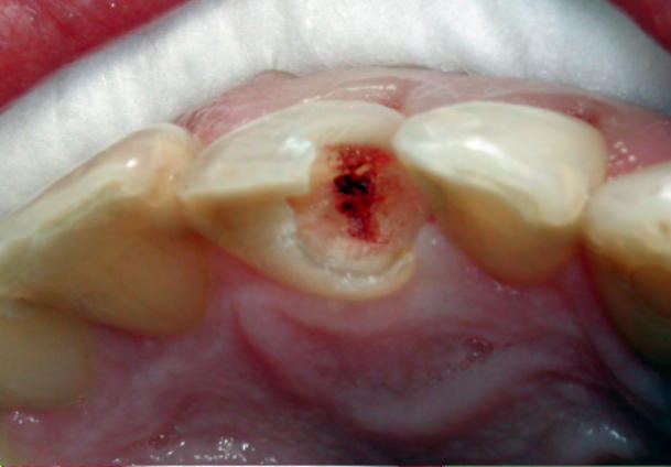
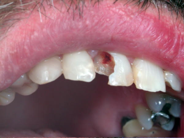
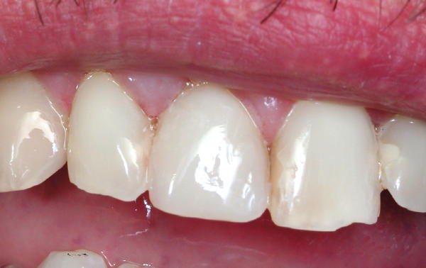
whitertthSpectatorWhat a day…saw this patient today and (someone call me an idiot for not having a preop) but u will get the drift. Removed large composite after using mark’s anaesthesia technique, exposed vital pulp, using the waterlase at .75 dryfor a few seconds i cauterized the nerve, etched , geristore over exposure, and bonded..no pain, not even a whimper… cauterized nerve in photo..post op to follow


Glenn van AsSpectatorMarvelous pictures Ron and really cool case.
I have had very very little success with this kind of treatment. Almost every carious exposure despite my attempts to maintain the pulp have ended in endo or a very calcified canal.
Hansen a while back provided some research on his technique and it had great success. I havent been able to duplicate things.
I like the final composite, very nicely done and thanks for posting the pics and NOPE I wont call you a dummy for no preop, it happens to me all the time!
Grin
Good stuff………
Glenn
whitertthSpectatorGlen,when has the nerve died on u ? I week after , a month 3 motnhs? I am gonna follow him every 2 weeks for a while….Monday I will post the radiograph…….
Having fun!!!
Glenn van AsSpectatorGreat question Ron…….the two cases that come to mind , I did the endos 2-3 months after the restorations were placed. One was a buccal and one a MO that come to mind.
I have two others that have had prolonged pulpal sensitivity that I am watching now and both are leaning towards the endo after the prolonged sensitivity to cold.
I have had two others where the sensitivity lessened. One was a 8 year old and the central ( traumatic fracture) discolored and vitality testing showed no pulp.
The other traumatic lateral calcified in on the pulp when the patient came back 2 years later because mom noticed the tooth getting darker and not matching the restoration……..pulp had calcified in which I thought was strange.
It just hasnt worked for me except on accidental exposures not chasing caries.
I would LOVE to hear what others are finding……in fact this is one area of using the laser that has disappointed me.
It may be something I am doing or it may be the pulp just cant respond despite our best efforts.
Who knows……..Mark? Bob? Ron?
What say ye
Robert GreggParticipantRon and Glenn–
Ron, nice case and photo presentation. From the amount of blood on the prep I would bet $$ this patient’s pulp is too injured to recover.
Yep. You’re right Glenn on the time frame. They tend to act up about 2-3 months after “vital” carious pulp capping.
The laser pulp caps that work are those where the exposure is limited in diameter to smaller then the diameter of the fiber-optic you are using, and/or the area of exposure can be isolated from the main trunk of the nerve/pulp (ie a pulp horn or a small axial pin-point exposure)
Carious/bacterial exposure: don’t expect much. Try if you want, but prepare the patient for future endo.
Bob
AnonymousGuestI know placing calcium hydroxide isn’t fashionable now but I continue to do so on exposures. I usually place the Calcium hydroxide and then ketac silver. I then etch and bond.
Thought this was interesting-Biocompatibility of a resin-modified glass-ionomer cement applied as pulp capping in human teeth.
do Nascimento AB, Fontana UF, Teixeira HM, Costa CA.Faculdade de Odonologia de Araraquara/UNESP, Departamento de Fisiologia e Patologia, Sao Paulo, Brazil.
PURPOSE: to evaluate the human pulp response following pulp capping with calcium hydroxide (CH, Group 1), and the resin-modified glass-ionomer Vitrebond (VIT, Group 2). MATERIALS AND METHODS: Intact teeth with no cavity preparation were used as control Group (ICG, Group 3). Buccal Class V cavities were prepared in 34 sound human premolars. After exposing the pulps, the pulp capping materials were applied and the cavities were filled using Clearfil Liner Bond 2 bonding agent and Z100 resin-based composite. The teeth were extracted after 5, 30, and from 120 to 300 days, fixed in 10% buffered formalin solution, and prepared according to routine histological techniques. 6-microm sections were stained with hematoxylin and eosin, Masson’s trichrome, or Brown & Brenn technique for bacterial observation. RESULTS: At 5 days, CH caused a large zone of coagulation necrosis. The mononuclear inflammatory reaction underneath the necrotic zone was slight to moderate. VIT caused a moderate to intense inflammatory pulp response with a large necrotic zone. A number of congested venules associated with plasma extravasation and neutrophilic infiltration was observed. Over time, only CH allowed pulp repair and complete dentin bridging around the pulp exposure site. VIT components displaced into the pulp tissue triggered a persistent inflammatory reaction which appeared to be associated with a lack of dentin bridge formation. After 30 days a few histological sections showed a number of bacteria on the lateral dentin walls. In these samples the pulp response was similar to those samples with no microleakage. VIT was more irritating to pulp tissue than CH, which allowed pulp repair associated with dentin bridge formation. These results suggested that VIT is not an appropriate dental material to be used in direct pulp capping for mechanically exposed human pulps.
PMID: 11763899 [PubMed – indexed for MEDLINE] [/quote]
dkimmelSpectatorIn cases like this would it be of any benefit to have the patient back and do a biostimulation tx ?
David
Glenn van AsSpectatorBob Gregg is the expert on LLLT ( Low level laser therapy) and biostimulation.
I am not a big guru in this area. I doubt it would improve the pulpitis much but I could be wrong.
Glenn
DoueckDentalSpectatorIf any of you are familiar with the name Gus Livaditis – it will bring you back over 20 years ago to the first lecture on the Maryland Bridge. Whatever the success rate you have had personally with the maryland bridge… It is clear that Gus is an innovator.
This time Gus has come up with a very predictable technique called “Vital Pulp therapy”. He has designed a special tip for a bipolar electrosurge unit (his unit with the tips is called the Dentstat – I paid about 񘡨 for the unit and 3 tips) Using this Denstat on a vital pulp exposure – no matter how large the exposure- I have been able to get predictable results in 90%+ of the cases with a 1 year post-op so far. Dr Gus Livatidis has been doing the procedure and has documented results for over 5 years. I am really excited about the results. There are two keys parts to this procedure
1. Durable Hemostasis – that’s why you need the bipolar electrosurge. The depth of penetration for the energy is very limited. The advantage of that over the laser energy is that the pulp tissue is not damaged. I have tried this technique with a diode laser and the waterlase… both modalities have too much energy to keep the nerve vital and healthy. With this technique the patient is comfortable right away and stays comfortable. I have used this for molars, and anteriors with excellent results.
2. The right pulp capping material – Dr. Livaditis has been using the meta-4 cement “MetaBond” – Personally I have found that material to be cumbersome to work with and I have been using J Morita’s version “M-Bond” which doesn’t need refrigeration and is very easy to work with.If you would like to discuss this please call me at 718-339-7982 Jacques Doueck Brooklyn NY
You can go on the website for Gus Livaditis
http://www.md-seminars.com/semtop_vitalpulp.phpFor complete documentation – go to the Journal of Prosthetic Dentistry and search for articles by Dr. Gus Livatidis.
Robert GreggParticipantHi David,
QUOTEIn cases like this would it be of any benefit to have the patient back and do a biostimulation tx ?Thanks, Glenn, but I don’t know how much of an LLLT expert I am compared to someone like Paul F. Bradley, BDS, MD, FDS, FR (no kidding).
To some degree, yes. It sould help with any symptoms and inflammation. And I don’t think it can hurt anything.
I think this may be a little too open for a full recovery, but that depends on things like the age of the patient, whether there is root apexification, etc.
Give it a try and let us know!
Bob
dkimmelSpectatorBob, Know of any good references on LLLT?
Thanks
David(Edited by dkimmel at 9:12 pm on Feb. 18, 2003)
Robert GreggParticipantYes. Yes, I do.
Bob
dkimmelSpectatorBob, Ok you got me on the hook!
David
Robert GreggParticipantDavid–Just funnin’ while I remembered where I put those references…..
Here’s a great one for everyone to start with:
1. Dr. Paul Bradley’s video from the 1999 ALD meeting in Palm Springs, “Current Status of LILT”. #039
Very informative, very well presented and entertaining.
Contact Joyco MultiMedia and ask for Bob or Joy (tell ’em I sent ya’) at P: 303-421-0093; Fax 303-403-9112
2. Low Level Laser Therapy by Tuner and Hode available from Dr. Larry Lytle of Lasers, Inc. 605-342-5669. Good book. Larry is also a great sourch of information.
3. The Science of Low Power Laser Therapy
by Russian researcher Tina Karu available at Amazon.com for ๳.95 (used to be around 趚).” target=”_blank”>http://www.amazon.com/exec….ooks
Karu’s book is by far the most scientific and also the most dry…….
Larry Lytle is usually at the ALD meetings and is always very fun to talk to. Ask him about “proud tissue”.
Bob
dkimmelSpectatorThanks Bob!
Sure will ask Larry about “proud tissue”!!
Sounds like getting some of my city friends to order Mountain Oysters at the local dinner!!
David -
AuthorPosts
