Forums › Erbium Lasers › General Erbium Discussion › A case where the scope and lasers helped
- This topic is empty.
-
AuthorPosts
-
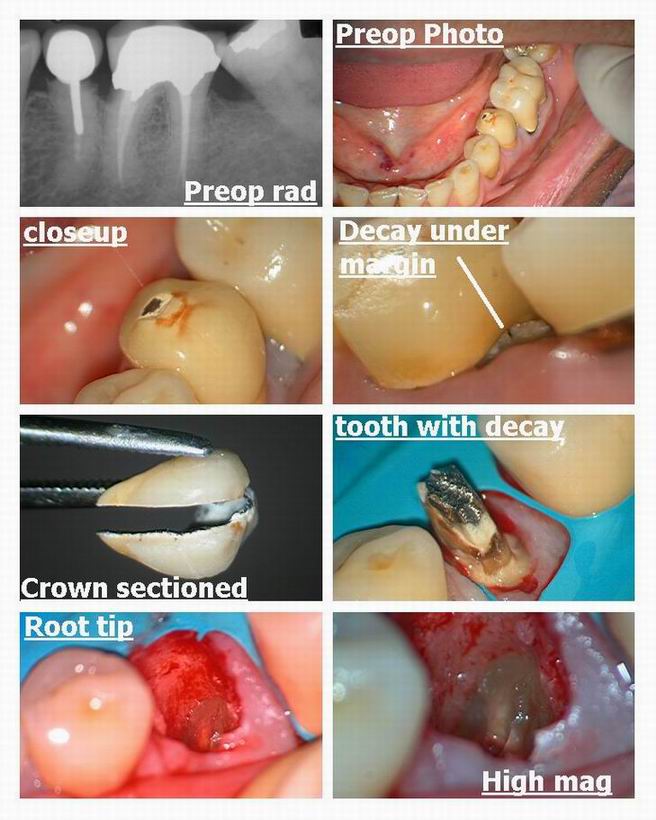
Glenn van AsSpectatorHi folks: Patient came in yesterday with this premolar on the lower left.
Rampant decay under the abutment, and note how that can be seen with the scope. Decision made to remove the tooth and the crown on the first molar and prep for a bridge.
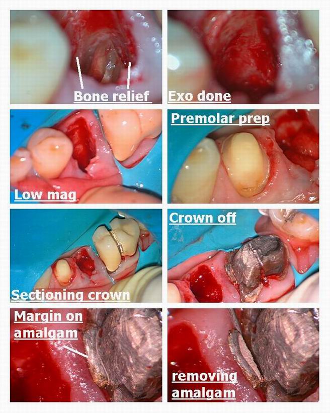
Premolar broke off but look how you can see the root tip at 10X power and drill around it with a small diamond and get it out. THe lighting and magnification really helped me get the root tip out
Then I removed the crown on the molar……..margin placed on amalgam in two spots.
Once the amalgam was out , no biologic width on the mesial and so we quickly used the erbium.
3 watts or so without water for the soft tissue and with water for the bone.
Argon laser to coagulate, and then a properly shaped temporary bridge for the temp.
Need to wait a little while to evaluate healing of the socket.
No flap, no referral to the specialist for perio surgery.
Easy.
Glenn


Glenn van AsSpectatorHere is the last photo of the temp bridge….late for work
Glenn
[img]https://www.laserdentistryforum.com/attachments/upload/Resize of Resize of Osseous recontouring with bridge pt 4_p4.JPG[/img]
SwpmnSpectatorNice use of the Er: YAG for osseous crown lengthening on mesial of molar.
Some non-LASER questions:
1) I noticed the endodontic treatment and mesial curvature towards apex of tooth #20. Atraumatic removal of root fragments such as these can be frustrating. Once you removed interproximal osseous on the mesial and distal with diamond, did you simply use an elevator to rotate out the root fragment?
2) Are you viewing the treatment field through your microscope while you are performing an extraction?
3) Excellent provisional bridge. Is that an auto-cure composite(e.g. Integrity or Luxatemp)?
Al
Glenn van AsSpectatorOh Allen you always ask the greatest questions.
I will be brief………I am starting a DVD production tomorrow and need to go read my manual for my new digital video camera that we are going to hook up to the scope.
1. This root was a bugger to get out. I broke it off one more time before I got it all out. IT was tough to get a bur down all that way. I like the small diamonds for troughing (needle nose sort of) around the roots but in this case it took a while to get it but I could see it the whole way down.
2. Yes I am viewing the treatment the whole time through the scope. The illumination and magnification are important for visibility . Remember flaps are for access and visibility and most of the time if I can get at the roots , I dont need to raise a flap . Like in this case.
I also find that the video output from the scope allows me to have the opportunity for my assistant to suction EXACTLY where I need it. She can see from the monitor positioned for her where to be an where to not be.She is an awesome assistant and is really a big help in tough ones like this where she is suctioning out of my way and right where the bleeding is. She gets a good idea of where to go from the macro view (she looks at the operating field ) and then fine tunes it by going to the micro view on the monitor.
She never stands for suctioning and I never have to grab the suction……..she is that good.
As for the provisional ……thanks. I use Luxatemp I think (sheesh I should know) but used a template made from the original teeth in a suck down shell. Placed gelfoam in the socket and then added composite to create an ovate pontic site.
Its one of those ones in a gun……..Luxatemp I think and then once adjusted we sandblast and place optiguard or a varnish overtop and cure it.
THat one actually wasnt one of my best.
Thanks Allen………you always make me want to post again.
Glenn
ASISpectatorHi Glenn,
Very nice handling of the tissue in extraction of premolar and prep of molar. Was the option of immediate implant to replace premolar not appealing to patient? Too bad the first premolar had to be involved.
Don’t mean to doubt your treatment planning. Just hate to see prepping of a virgin tooth.
Andrew
Glenn van AsSpectatorHi Andrew………no offence taken.
good point about the implant…hadnt really thought about it to tell you the truth.
The crown came off on the molar a while back and the margin was on amalgam my associate mentioned to me.
I knew that I had to replace that crown as well as extracting the premolar.
An implant was a definite alternative and one that I should have thought more about. I dont know if this patient would have taken the chance as he is very conservative in his approach only going for dental plan coverage.
I will next time take your input and see if the patient wants implants, but I was kinda thrown off because I didnt originally see the gentleman but my associate did and scheduled him for crown lengthening.
You know what I got another crown lengthening diagnosed today with a fracture premolar…….there are alot of them if you go looking for them and dont ignore them………..amazing and easy to do with the laser.
Thanks Andrew…….I am not to big to recieve constructive criticism for the cases I post……..both you and Allen did that and I will be better prepared in the future…….
Thanks alot for the input.
Glenn
AnonymousGuestThought I’d take 1 last look before leaving .
Glenn, does Hoya make long tips for your Er?
In cases where I’m pretty sure the tooth will crunch I’ve been using a long (12mm perio?) tip to remove bone interproximally. In this case I would remove enough interproximal bone on the mesial to allow that tooth to rotate out toward the mesial. I would also remove just enough on the distal to get a straight elevator in and know I’m fulcruming off bone and not adjacent tooth. With the straight elevator on the distal I would use a clockwise rotation along with downward movement to elevate the tooth. Sometimes I’ll have to also just rotate the elevator on the mesial also.
I’ve done many this way now, and so far the patients all have seem to have got along with just ibuprofen or no pain meds. I also feel more comfortable doing these when I know I won’t have to go in with the high speed and if I do still break the tooth I can ‘paint’ the gingiva with the laser to eliminate that bleeding and help improve visibility.
The pictures are awesome and your patient is fortunate to have someone like you doing the work.Thanks again for ‘filling ‘ in as admin.
Ron -
AuthorPosts
