Forums › Laser Treatment Tips and Techniques › Hard Tissue Procedures › Apicoectomy
- This topic is empty.
-
AuthorPosts
-
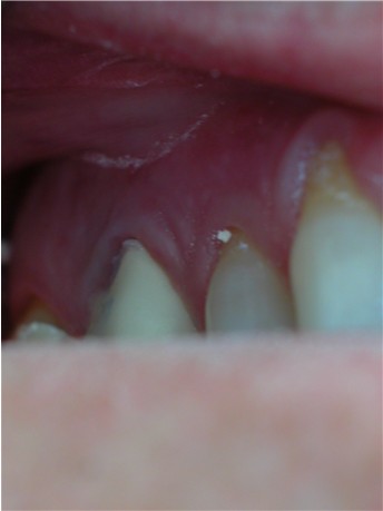
kellyjblodgettdmdSpectatorI wanted to share an apico I did last week. The patient had fractured #2 and was coming in as a new patient wanting Tx. A radiolucency was noted at the MB root tip. Tooth #3 had received RCT and also retreatment, last treated approx. 3 years ago. The patient wanted all necessary Tx performed in one visit.
For the apico I:
1) Marked the line for the incision w/ the Er,Cr:YSGG @ 0.5W – 0/0 for air/water
2) Made incision w/1.0W @ 14/8
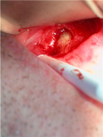
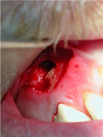
3) Removed buccal bone revealing root tip and apical pathology 3.0W @ 60/60
4) Removed 2mm off of root w/ 3.5W @ 65/60 and removed pathological tissue w/ 2.0W @ 65/60.
I didn’t get a picture of the apical filling or sutures. Sorry. When I called the patient that night he reported mild tenderness, but said overall he was doing pretty well. I felt that using the Er laser for this was a far better option than with knifes and burs. The bleeding was minimal. I think he will heal nicely and finally be able to chew on this tooth.
kellyjblodgettdmdSpectatorSorry – somehow I screwed up the attachments. Here are the four pictures:


(Edited” target=”_blank”>https://www.laserdentistryforum.com/attachments/upload/Joe2.jpg%5B/img%5D


(Edited by kellyjblodgettdmd at 9:27 pm on May 28, 2003)
SwpmnSpectatorInteresting, thanks for sharing. Hadn’t really seen a documented laser apicoectomy.
After you made your Erbium incision did that alveolar mucosa just peel right off or did you have to elevate the mucosa with an instrument?
I haven’t done any “flapped” laser osseous procedures like yours although I’ve done a handful of these “closed” crown lengthenings. When we’ve got a flap reflected do we know what the risk is of getting an air emphysema with the Erbium? Would it be comparable to the risk of using a non-surgical high speed handpiece?
Thanks,
Al
kellyjblodgettdmdSpectatorThanks Al,
With regards to the flap, it took minor, light pressure elevation, but came up relatively easily.
With regards to air emphysema, I’m not terribly concerned although it is always a consideration. I have never used anything in the past except standard high-speed hand pieces for surgery. Now, I’m not doing LeFort II’s or anything, but I’ve done my fair share of sectioning teeth, 3rd molars, and alveoplasty with high-speeds in the past and haven’t had any issues.
The beauty of the Erbiums is that you can easily control the amount of water and air you feel comfortable using. I feel that as long as I have the tissue properly reflected, there is little risk. After seeing so many surgical slides at the World Clinical Laser Institute with Doctors using the Waterlase, I felt very comfortable using their suggested settings as a guide for my more “conservative” surgeries.
Just my two cents. Thanks for the reply.
Kelly
2thlaserSpectatorKelly,
Great case, and hopefully the result will be too. Thanks for sharing. Did you use a T tip? for the incision?
Mark
kellyjblodgettdmdSpectatorThanks, Mark. I did use a T-tip for the incision. For the bone removal and root-tip sectioning, I used a G-6.
I must say, the incision was so easy to suture. I should have the post-op pictures in ~ 2 weeks.
ASISpectatorHi Kelly,
Nice work. Great almost bloodless field to work in, isn’t it?
What kind of material did you use for the apical fill?
Andrew
kellyjblodgettdmdSpectatorThanks, Andrew. I packed some MTA into the apical prep. The specialized carriers made by Dentsply make it easy to carry.
Kelly
whitertthSpectatorGreat stuff….. Keep up the good work
webpiterSpectatorYour teeth are held in place by roots that extend into your jawbone. Front teeth usually have one root. Other teeth, such as your premolars and molars, have two or more roots. The tip or end of each root is called the apex. Nerves and blood vessels enter the tooth through the apex. They travel through a canal inside the root, and into the pulp chamber. This chamber is inside the crown (the part of the tooth you can see in your mouth).
During root canal treatment, your dentist cleans the canals using special instruments called files. Inflamed or infected tissue is removed. An apicoectomy may be needed when an infection develops or won’t go away after root canal treatment or re-treatment.
-
AuthorPosts
