Forums › Laser Treatment Tips and Techniques › Hard Tissue Procedures › Cracked tooth
- This topic is empty.
-
AuthorPosts
-
2thlaserSpectatorRemember the case on my uncle? Well here’s an interesting one on my aunt. She had chronically been complaining of a “bump” on the inside of her gum for some time, while she was at home in PA. I had her see an endodontist, who then did an apico, with no resolve. Next, he performed “flap surgery” where he thought some peiodontal pocketing was occurring, he did the procedure 2 times in a 2 month span, not finding anything, not a crack, nothing. I asked my aunt if she remembers him using magnification, either loupes (showed her mine), or a microscope, and she couldn’t remember…anyhow, here is the preop radiographs…
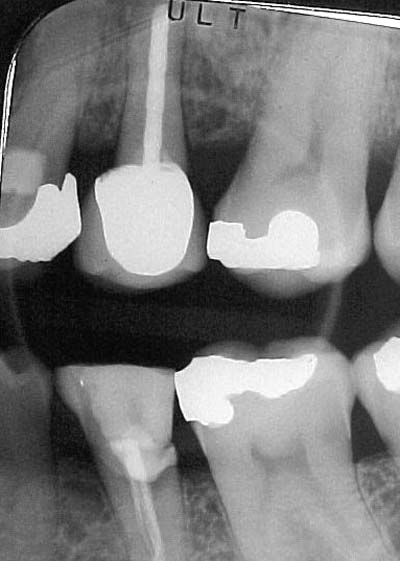
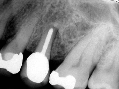
Sorry, no preop photo’s, I was VERY short on time, UNTIL I removed the crown…and here is what we saw IMMEDIATELY after removing the crown with a great white bur…..
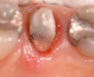
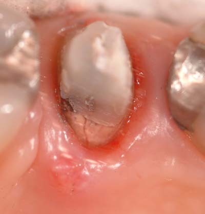
I took slightly different angles to show the fracture from the post. It was a parapost, with a composite build up. The tooth had normal occlusion, no atraumatic forces..just a chronic fracture of the root. I really think that with magnification, a surgeon would have saw the fracture upon flapping…anyone disagree? Here is the tooth extracted, note the length of the fracture, and where the bone had been destroyed by the bacteria.
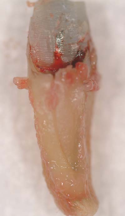
I hope you guys don’t mind me posting this. I thought it was interesting. Glenn, THIS is a good reason for magnification, don’t you think? I think a scope here would have been very beneficial.
Today, we prepped her for a bridge, she didn’t want to wait for implants…we move on!
Thanks,
Mark
Glenn van AsSpectatorMark: absolutely cool……..I will post a collage of cracks we see with the scope. The two things you notice right away are cracks everywhere and decay.
Dr. David Clark from Washington state just has completed a cool article on categorizing cracks based on 16X mag and it is very very thought provoking. I am peer reviewing it for the Journal of Esthetic Dentistry and I think that you are going to see alot of stuff coming out that will advance cracks and their treatment before end stage disaster in the future, and most of this will be driven by high magnification technologies such as the scope.
If you think about it all our treatment for cracks is symptom driven, wouldnt it be cool to treat these prior to them becoming sore.
Just a thought for you……….Great pics and I will post a collage of cracks if you want to see them……..
All shot at high mag.
Glenn
I will wait to see if people want to see them because they really arent “laser dentistry” per say although I have treated some of the non vertical kind with the laser before finishing the buildup.
Cool pics ……..FUji S2??
Glenn
PS kudos for getting it out in one piece.
2thlaserSpectatorThanks Glenn…I would LOVE to see your pictures. More the merrier, I want to learn buddy!
Yes, these are all from the S-2. It is such a great camera. Very easy to edit. Also, thanks for the advice on posting xrays…got it down I think.
I just wanted to show what posts and pins do. I have another picture of a pin related crack, BUT with the scope I am sure you have a myriad of them. I would love to see them.
Thanks,
Mark
Glenn van AsSpectatorOk…..here are just a few I have hanging around…..
Glenn
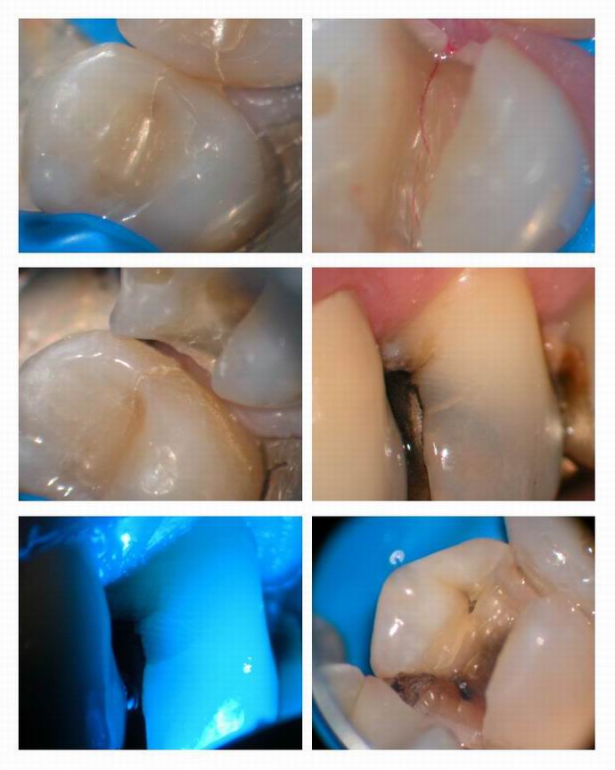
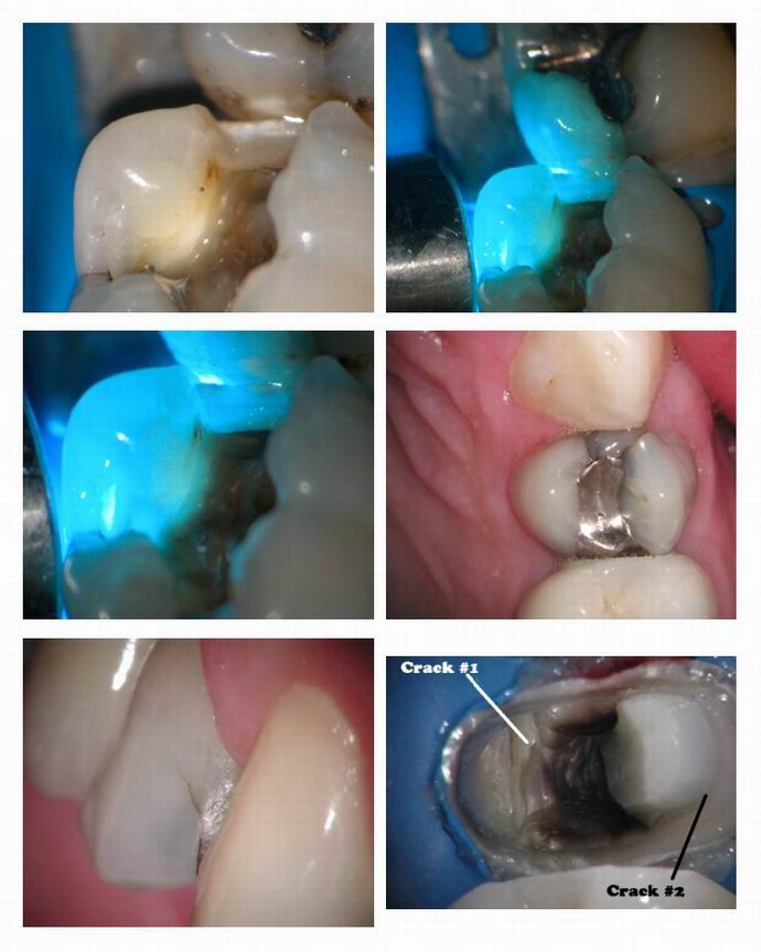
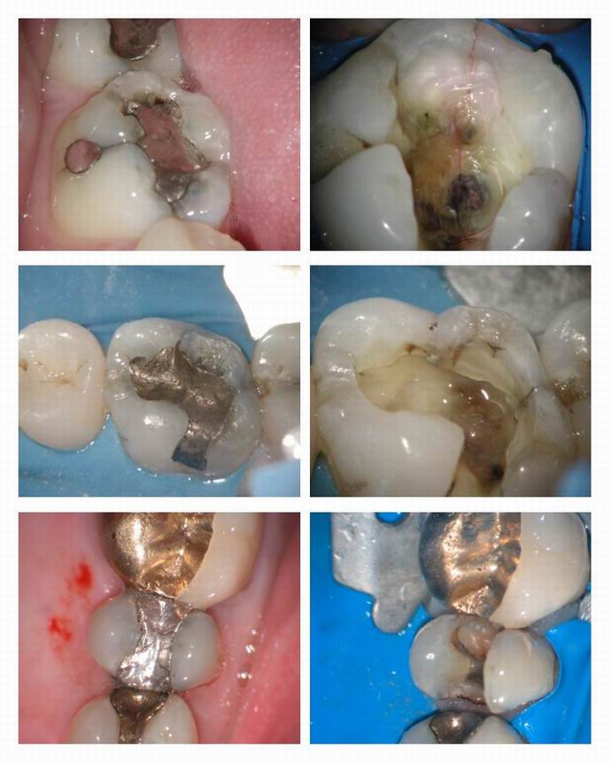
Glenn van AsSpectatorHere are just a few more…….gotta go and do some lectures…..
Cya
Glenn
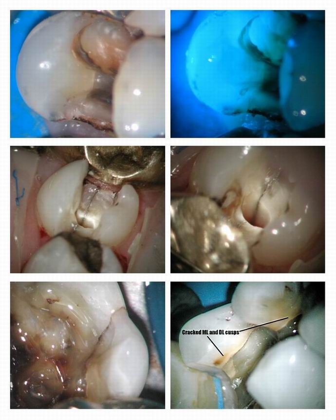
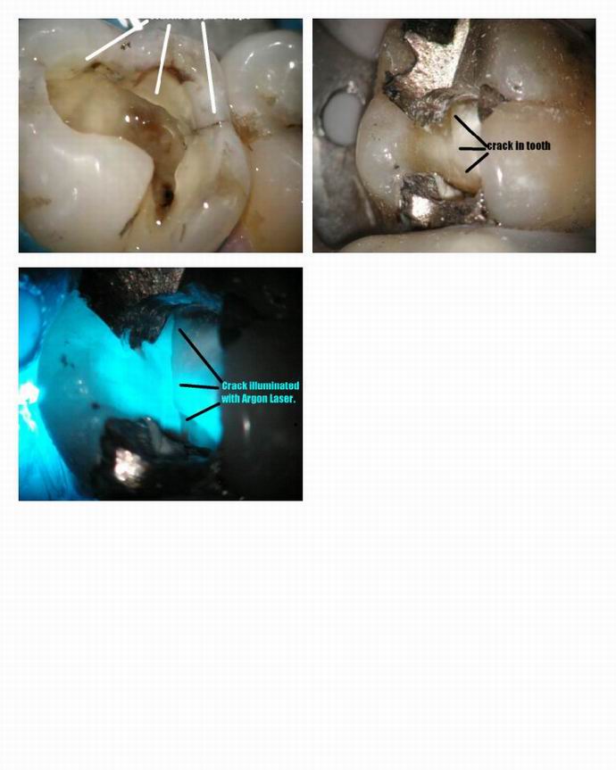
2thlaserSpectatorHoly Cow, These are GREAT images Glenn! Thanks so much. What a great adjunct a scope is. Maybe Ron ought to think of having a magnification thread…?
Mark
Glenn van AsSpectatorSounds great to me………I am not big on the DT one as I get ridiculed constantly about it. PErhaps after the Dentistry Today article comes out in June then there will be more interest. Its a coming technology and it is interesting to see all the cracks.
I see them each day but to be honest with you have never thought about them the way David Clark has but I tell you it is amazing how many I see under Occlusal amalgams.
If Ron wants a magnfication forum for lasers or whatever , I am happy to help out there.
Glenn
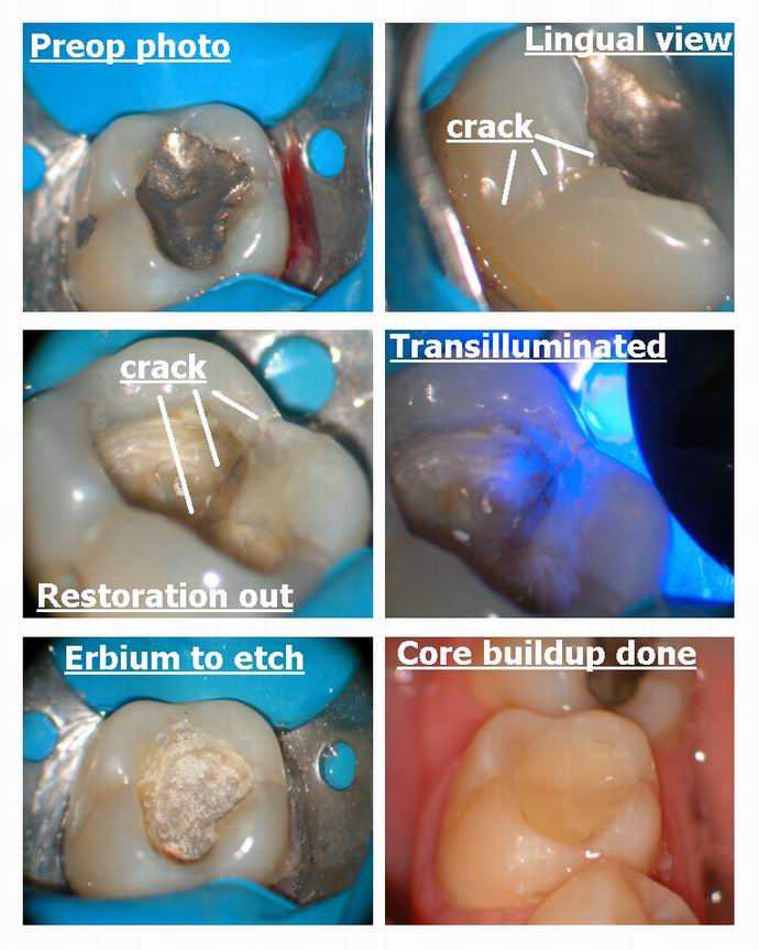
Glenn van AsSpectatorHere is a classic case for you Mark. A lady with an occlusal amalgam that is sore to chewing. You think the dental plan will pay for a crown……hahaha.
Open it up and take photos for the plan, they wont not approve it then when I show them the crack and build it up. I see this at least once per day I would guess. Usually first molars and wide restorations place 15 -20 years previous. Now I dont want to argue if the amalgam expansion does it, the bur, bruxism…..it happens that is all and patients want answers.
Kinda cool when you can pinpoint EXACTLY where the crack is……photograph it and then show the patient the solution (crown +/- endo).
Glenn
ASISpectatorHi Mark & Glenn,
Nice documentation of cracks in teeth. I see them all the time even without any magnification. Hoping to get into this soon though.
Are you using scopes as well, Mark? The images look magnified.
Andrew
dkimmelSpectatorGreat posting on cracks!
Glenn, I am not getting a scope! I am not getting a scope! I am not getting a scope! Well not just yet!
So much to buy to improve dentistry! So hard to budget and stay within those limits!
David
Glenn van AsSpectatorI couldnt agree with you more David. It is getting exhausting staying up with technology but I will say this that the scope is really valuable in the practice as I honestly use it for every single patient for almost 100% of the procedures. I might not use it for anesthetic or rubber dam placement and sometimes not for the placement of the matrix band in the maxilla but almost for everything else.
For lasers it really helps alot with seeing what you are doing. Magnification is essential in my humble opinion to do a great job with the laser. Scopes are an added bonus, I truly believe in my heart that both the magnification and the illumination both help with making the dentistry better.
We all still make mistakes….I had an accidental exposure yesterday on a little 5 year old who wouldnt hold still. I was trying to go fast with a 400 micron tip and nicked the pulp……..silly on my part but accidents still happen but with far less frequency with the scope.
ALl the best and thanks David………your comments do fall on deaf ears (or in this case blind eyes!!)
Cya
Glenn
AnonymousGuestQUOTEQuote: from dkimmel on 11:08 pm on May 2, 2003
Great posting on cracks!
Glenn, I am not getting a scope! I am not getting a scope! I am not getting a scope! Well not just yet!
So much to buy to improve dentistry! So hard to budget and stay within those limits!
DavidDon’t forget the perioscopy
and the Tek-Scan
and the…..
and the…
and the..
dkimmelSpectatorRon, I have them coming Friday to demo the perioscope!!
David
Robert Gregg DDSSpectatorHi All,
I don’t have a lot of technology (I don’t think). I have a lot of lasers–and a Global scope.
Not much else. No AA, no digital x-rays.
I like the scope as it aids me in clinical practice, which is how I judge my purchases.
Bob
-
AuthorPosts
