Forums › Erbium Lasers › General Erbium Discussion › Lasers and cracks
- This topic is empty.
-
AuthorPosts
-
ASISpectatorThanks, Glenn.
The voice of experience is heard loud and clear. For your scoped eyes have seen much that many unaided eyes or lesser aided eyes have not been able to.
Thanks again.
Andrew
Lee AllenSpectatorGlenn,
The followup on my patient with the bite sensitivity: I took Mark’s advice and removed all of the class I composite and found that indeed there was a crack under the distobuccal cusp adjacent to the area treated last. This was a tooth that had been restored with all bur, was 4 mm deep and off center to the distobuccal making that side thin. Prior to anesthetic, it did test postitive to the Tooth Sleuth in the same area.
This will be interesting to follow since the patient did not want to have a recommended crown (I use an intraoral camera to show patients their tooth–poor mans version of the scope), so a composite replacing the cusp was placed over a GI base. I find less contraction forces at work when I place a big base. I expect the patient to be back for the crown.
Thanks to all for the help in sorting out this one.
Lee
2thlaserSpectatorLee,
You are welcome from my end. You are such a good dentist…for those who haven’t met him, trust me, he is! One other thing I do when I find those cracks, I lase into them, making sure that there is NO bacteria in them as much as possible. I also believe that this area is strengthend a bit by the restorative materials upon setting. Just a thought. Anyone else with thoughts on that? Teach me!….
Mark
SwpmnSpectatorMark I think you are exactly right and I do the same thing – lase right into the crack then use my bonding agent and flowable comp prior to my restorative composite.
Al
Lee AllenSpectatorGlenn,
The immediate post op results on my patient with the internally cracked DB cusp: 3 days PO and she is without pain even to temp and chewing. Considers it a miracle and so do I. Certainly new territory for me.I am wondering if perhaps some of the CTS (cracked tooth syndrome) that I diagnose could be treated by composite restoration. In this case I did cover all the visible cracks with composite, troughing the outside and inside of the crack to fill with flowable.
Also, I am interested in the “lasing” of the crack that Allen and Mark are doing. Sounds like it is not defocused but is entered from the inside of the prep without penetrating to the outside surface. Does the presence of stain signify anything with regard to sucess, to long presence of the crack, to bacterial activity? If there is not staining, do you still lase the crack and seal with flowable internally prior to the rest of the compostie placement? Or in the end are the patient’s symptoms the driving force behind the decision to lase or not to lase.
Now I see why a picture is so valuable. 1000 word thing.
jetsfanSpectatorGlen,
Today , for the first time I actually made a mental note of what I saw upon entering a tooth with a diamond ,using my 4x magnification. I actually could see a “microcrack form as soon as the diamond entered the tooth. I now realize that I have seen this often without thinking twice about it.
2thlaserSpectatorNow you know why I don’t use a burr. I try SO hard not to crack these teeth anymore. Thanks for sharing Jetsfan!
Mark
jetsfanSpectatorI noticed something else yesterday, which I have seen before but never thought twice about: A patient has a OL amalgam on a max first molar and there is a crack which runs fron the filling gingivally. It is stained dark.
I always believed that it was the large filling causing the crack in the tooth. Now I’m not so sure. Perhaps it was the metal bur in the drill that caused the microcrack and it was stained over time by the amalgam.
Graeme MilicichSpectatorGlen
Looking at the tooth, I believe youa re looking at Occlusal effect fracturing. By that I mean, by removing the occlusal enamel, you remove the occlusal cross bracing and destabilize the peripheral rim of enamel. Compressive loading on the enamel rim leads to flexing which cracks the enamel in the area of the marginal ridge.Cheers
Graeme
Dave RodrickSpectatorGraeme,
What do you think about cleaning the dentinal surface and the cracks with a laser and then placing GI base after a 5 sec rinse with cavity cleanser. Then adding cross bracing (buccolingually) with ribbond and fill around with a resin filling as a differential diagnosis. After adjusting occlusion and waiting a couple weeks check on symptomology and decide to do endo if symptoms persist?
Dave
Graeme MilicichSpectatorDave
When you look closely at Glen’s photo, there are no dentin cracks.
What youa re suggesting would be a conservative way of assessing the problem. THe need is to stabilize any dentin cracks, either by disecting them out or reducing and overlaying the cusps.
Cheers
Glenn van AsSpectatorHi Graeme: I really respect your opinions on alot of microdentistry but in this case I will disagree with you.
I dont know if you are using the microscope at all but I know that when I removed the amalgam the reason I removed the cracks is that the crack was right through to the previous amalgam.
I see these cracks ALL the time get wider and then become a V with a piece of tooth structure broken down.
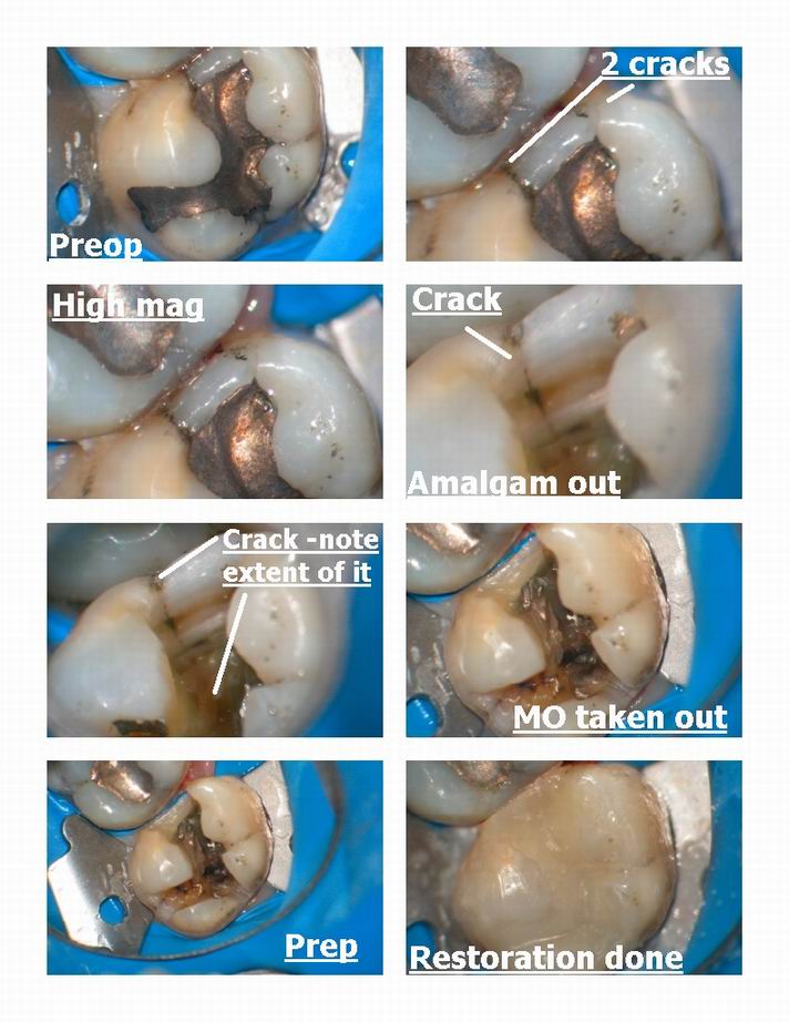
Pairs of cracks I remove especially when they are in the marginal ridges. Dr. David Clark is coming out with a new categorization of types of cracks as seen under the scope and how to treat them. Although these marginal cracks rarely do break into the pulp , unless they are singular, I am not a big fan of leaving them there in the marginal ridge if after I remove the amalgam that they are present on the internal aspect of the prep.
I realize the photos dont show that but for me the future breakdown of the tooth goes beyond the integral strength of the marginal ridge (which I do believe is very important and why so often you can restore the case conservatively without destroying the marginal ridge).
I will look to see if I have photos
1. Crack in this case or another which is present after the amalgam is removed.
2. Conservative nature of preparing ONLY the interproximal decay (with the scope) to avoid breaking down the marginal ridge.I learned number 2 from your postings before and try not to destroy it BUT in the case where the marginal cracks are also visible on the internal aspect of the preps , they are removed in my practice.
Just my two cents worth but if we want to get into a discussion of cracks , I have TONS of photos of them, more than anyone can imagine and I can slowly start to discuss some of the new criteria we are discussing for the treatment of cracks (BEFORE THEY BECOME SYMPTOMATIC- now wouldnt that be cool huh!!)
Great post Graeme,
Glenn
Glenn van AsSpectatorGraeme: The area of cracks is absolutely so poorly discussed because we dont have scopes and we cant see how prevalent they are.
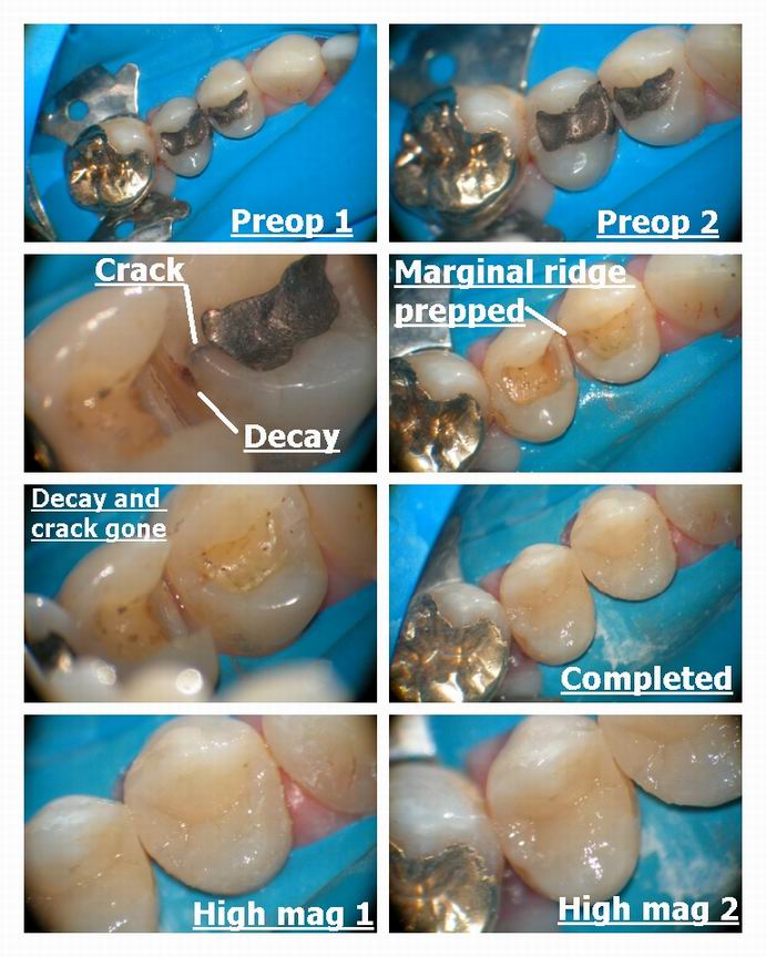
One thing that we find a TON is decay under these cracks interproximally.
HEre is an example of that and how I treated it.
Glenn

Glenn van AsSpectatorHere is another similar case where the amalgam was removed and look how far the crack went on the marginal ridge into the tooth.
Glenn

ASISpectatorHi Glenn,
Great stuff, Glenn.
These are quite the usual situation from crack at the marginal ridge area. Now with the scope, I detect them so much better and they are so much more evident as well.
Miss seeing your photos, man.
Andrew
-
AuthorPosts
