Forums › Erbium Lasers › General Erbium Discussion › Lasers and cracks
- This topic is empty.
-
AuthorPosts
-
Glenn van AsSpectatorThanks Andrew: great to be back . Off to Calgary for a big lecture on Thursday and frantically finishing that off.
With the scope you can see whether they are into the dentin or still in the enamel.
I only remove the ones that are big.
Thanks for the great comments.
Talk to you soon.
Glenn
Graeme MilicichSpectatorGlen
I have been using a scope since 1998.I believe the two latest posts show two different presentations of fracturing.
This is really hard to try and get across in words but I will give it a go.
The first one doesn’t exhibit any dentin fracturing, just enamel fracturing, with caries in the underlying dentin.
This is what we are calling occlusal effect caries. This is caused by compressive load on the cusp distorting the peripheral rim of enamel, which cracks the enamel, then caries establishes at the EDJ and grows into the dentin and outward through the wall of the cracked enamel.
I wrote an article on this concept that was published in Sept 2000 in PPAD
Milicich G, Rainey JT. Clinical presentations of stress distribution in teeth and the significance in operative dentistry. Pract Periodontics Aesthet Dent 2000 Sep;12(7):695-700
you can access it online through my website
<a href="http://www.advancedental-ltd.com/index.htmand” target=”_blank”>http://www.advancedental-ltd.com/index.htm
and the article is on the resources page. It is a link to a monatage media pdf file so you get to see it all in colour.
This will be the easiest way to try and explain the concept of occlusal effect caries associateed with compressive fracturing of the peripheral rim enamel.
The other tooth (the Molar) exhibits the more common tensile load fracturing. This does extend into the dentin due to dentin’s inability to handle tensile loading. The tooth is set up biomechanically to deal with compressive loading only Excessive tensile loads can destroy a virgin tooth, let alone one we have gone and removed all the occlusal cross bracing structures from.
Have a look at the article and tell me what you think.
The intersting thing with occlusal effect caries is the dentine caries is very difficult to detect in its early stages because it is so focused around the crack that it doesn’t involve enough bucco-lingual width to be detectable on Xray. We are playing around with some new software called Logicon that uses a digital analysis using 256 shades of grey instead of what the human eye can detect (10-30 shades)
Cheers
Graeme MilicichSpectatorHelp
How do you get images into the post. I uploaded some onto the site, but can’t find them to put into this post.
I tried to upload them a second time, but I can’t because they are already somewhere on the site.
Cheers
Dave RodrickSpectatorGraeme,
I understand cracking occuring because of occlusal loading but how many of these cracks, in moderate to large amalgams, are due to the old amalgam swelling with time. I once read an article about “water sorption” and the ability of amalgam to swell as it gets older and act as a wedge to cause fracture lines on marginal ridge and then subsequent caries development. I have some pictures of old amalgam looking like its swelling out of the preparation.
Dave
SwpmnSpectatorQUOTEHow do you get images into the post. I uploaded some onto the site, but can’t find them to put into this post.
I tried to upload them a second time, but I can’t because they are already somewhere on the site.Graeme:
After you get the message “Your images were successfully uploaded!”, make sure you click each image individually so that they will appear in your post. This may explain how you lost the images.
Enjoyed your lecture on occlusal effect caries/peripheral rim theory at WCLI in Atlantic City. Also, your technique of scaling erbium prepared enamel margins has been most useful in reducing the “white line effect”!!!
Al
Graeme MilicichSpectatorHere is what I am trying to describe. When you cut out the occlusal cross bracing, lateral compressive loads (circled areas with the load direction indicated by the arrows), casues the tooth to distort. The distortion is like when you squeeze a baked bean can that has had the top cut open. (when you squeezr the can, it becomes ovate, with the apex of the distortion about 90 degrees around the can from where the compression is being applied)
The same thing happens to teeth that have had their lid cut out. Dentin is compressible, enamel is not. Therfore it will crack when exposed to the tensile force associated with the underlying dentin bending.Walker BN, Makinson OF, Peters MC Enamel cracks. The role of enamel lamellae in caries initiation
Aust Dent J 1998 Apr;43(2):110-6 Department of Dentistry, University of Adelaide, South Australia.This article showed the development of careis that starts at teh EDJ underneath these unstable enamel fractures. These cracks need to be differentiated from ordinary enamel lamellae which are stable and generally of no long term significance.
Wang, R Z Weiner, S. Strain Structure in human teeth using Moiré fringes Journal of Biomechanics 1998 Feb:31(2):135-141. Strain distribution within the tooth is related to its structure. Enamel acts as a stress distributor, transferring the load vertically to the root and horizontally into the dentine of the crown.
This article points towards how the tooth functions under load and lead us to the concept I am trying to describe here.
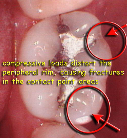
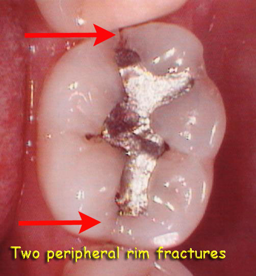
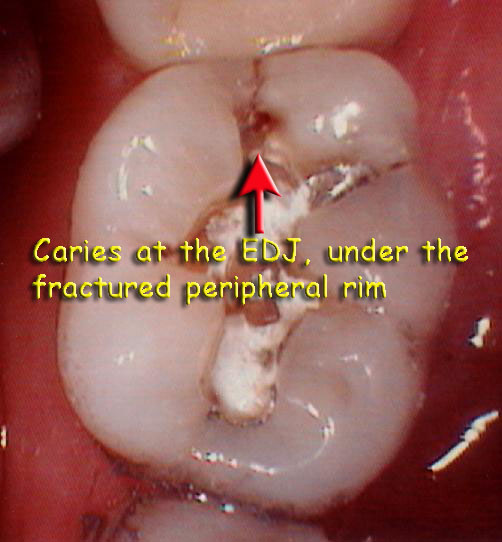
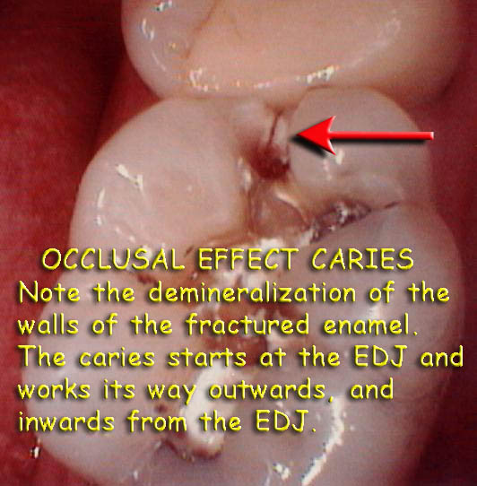
Hope these images help with understanding this concept. This is compressive loading of the peripheral rim leading to fracturing of the enamel only.(ie lateral loading from the buccal face is pushing the cusp ONTO the tooth)
Tensile loading of cusps (ie lateral loading is trying to push the cusp off the tooth) exposes the underlying dentin to tensile forces rather than compressive forces, leading to fracturing of the enamel and dentin.
Hope this makes sense
Cheers
(Edited by Graeme Milicich at 1:53 pm on Sep. 17, 2003)
Graeme MilicichSpectatorDave
Amalgam can “grow” in that it undergoes plastic creep, which leads to the amalgam “growing” out of the cavity.I am sure some fractures we see can eba tributed to pressure from distorting amalgam. another potential source of this pressure is continuing buildup of corrosion products slowly jacking the cusps away from the amalgam.
However ther a re many more fractures that occur that cannot be accounted for by this mechanism.
I tried to upload some differnet type of fractuer images but all I got this time were boxes with crosses in them!!!
Cheers
(Edited by Graeme Milicich at 2:07 pm on Sep. 17, 2003)
Graeme MilicichSpectatorAnother try!! I had a # in the file names and I think that screwed everything up.
How many times do you see this type of fracture. The peripheral rim enamel has delaminated from the occlusal enamel (yes they are different in the way they function)
What is happening here is is the stability of the tooth has been destroyed by the cutting of a G.V.Black cavity. The presence of the amalgam contributes absolutely nothing to how the tooth is now functioning at the biomechanical level. the cavity may as well be empty as far as the tooth is concerned at the functional level.
Lateral compressive force (highlighted with bite paper (inside red circle) compresses the buccal peripheral rim enamel onto the tooth. Enamel is very strong in compression, but weak in tension.
The underlying dentin distorts under compressive load, and the overlying enamel begins to delaminate from the occlusal enamel, creating this “occulsal abfraction groove”. Eventually you see total loss of the occlusal enamel section down to the dentin, with the peripheral rim left standing proud. The last image is the contralateral tooth of the tooth in the first two images. The failure has just progressed to the next stage.These only happen because we cut out the cross bracing structures like the sub-occlusal oblique transverse ridge in lower molars.
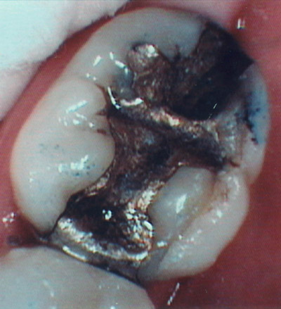
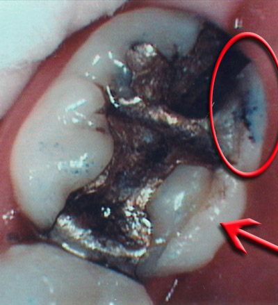
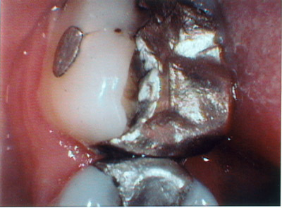
These fractures are not directly attributable to the presence of the amalgam, but to what we did to the tooth, cutting it to pieces to place an amalgam.
Cheers
ASISpectatorHi Graeme,
Thanks for your thoughtful presentation and photos.
GV Black cavity prep design did not truly take into consideration of longevity of the tooth structure in function. At his time, edentulism was at a greater incidence with age.
Teeth weren’t counted on to remain in function for as long as they are expected now. The dental education curriculum needs to evolve with contemporary dentistry.
Andrew
Glenn van AsSpectatorThanks Graeme: nice pics that show your thinking well. I do see alot of these types of cracks and it is interesting to think that the crack may come first then the decay. We would think it was the other way huh.
I still will remove the cracks when the proceed into the dentin on the marginal ridge because I do fear that the crack will lead to decay with time. It is a risk to the tooth and I agree weakens it but the crack is a sign that the tooth is already weakened.
I will post some pics of how I am able to limit my preps to the marginal interproximal surface in many cases with the power of the microscope.
I think that I would like to speak at the next WCM meeting on the value of microscopes for microdentistry. If pictures and descriptions like you provided are present , it would be a wonderful learning experience.
Sorry for the brevity , but rushing out the door to speak on high technology in Calgary.
Will be back on Friday.
Cheers
Glenn
Graeme MilicichSpectatorQUOTEQuote: from Swpmn on 9:49 am on Sep. 17, 2003Quote:Graeme:After you get the message “Your images were successfully uploaded!”, make sure you click each image individually so that they will appear in your post. This may explain how you lost the images.
Enjoyed your lecture on occlusal effect caries/peripheral rim theory at WCLI in Atlantic City. Also, your technique of scaling erbium prepared enamel margins has been most useful in reducing the “white line effect”!!!
Al
Al
In the early days I was getting frustrated with having yo deal wth the white spots and startes dinking around with them and stumbled onto the trick.I don’t get many now becuase I am more adept at using teh laser, but even now, they are impossible to avoid on some teeth. I think it has something to do with the presence of aprismatic enamel or hypermineralized enamel on the extreme outer surface of enamel because it is a surface phenomenon. You don’t see it happening in the deeper layers of the enamel.
Cheers
-
AuthorPosts
