Forums › Erbium Lasers › General Erbium Discussion › Mucocoele removal
- This topic is empty.
-
AuthorPosts
-
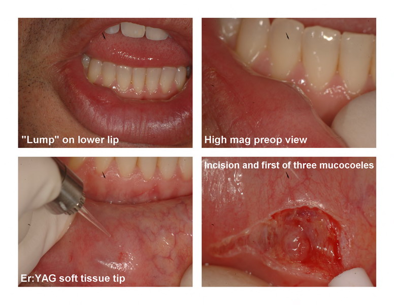
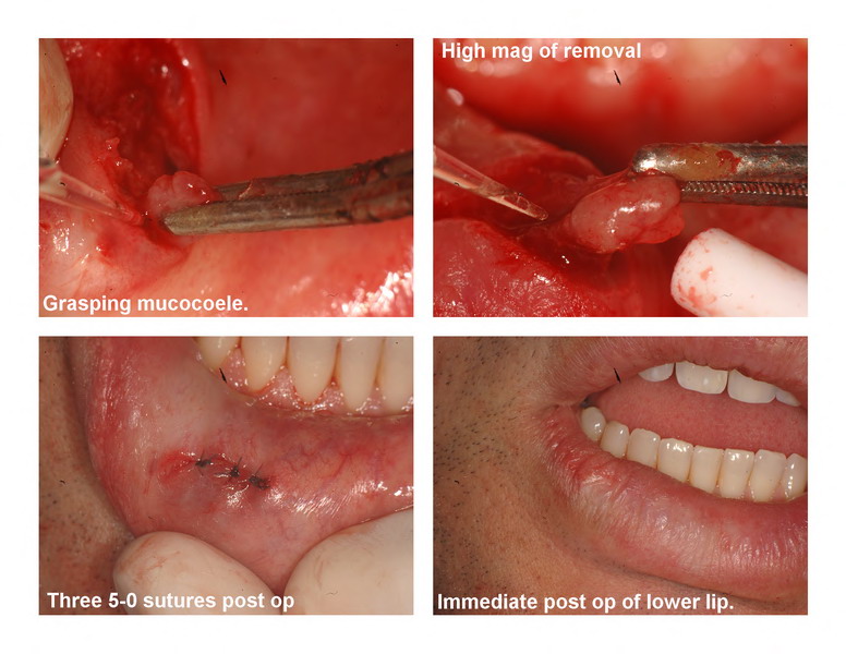
Glenn van AsSpectatorHi Folks: Here is a case that was completed during a hands on in our office on Friday.
THe patient was a mid thirties year old male who notice a lump in his lower right lip that grew and shrank over time but lately was bigger and becoming noticeable when he smiled. Palpation revealed a fluctuant lump which required removal. Suspicion of a mucocoele was my diagnosis.
I used the Er:YAG hard tissue laser and made an incision with the DeLight at 30Hz and 60 mj (1.8 watts) without water. Anesthetic was placed (20% of one carpule) and EMLA used before the anesthetic.
I was surprised at how right away the first mucocoele popped into view. I grasped it with a hemostat and easily dissected it away from the lip. Using the soft tissue tip to dissect around 2 more similarly enlarged glands we closed with 3 sutures (6-0) and will see the patient later on this week.
Quick and the visibility was excellent without much bleeding which really made it easy to see where the offending enlarged minor salivary glands were located. Oh sure a blade works well but the attendees mentioned how much more bleeding they have with blades and how much more difficult it is to dissect around the mucocoele it is with a scalpel.
Just wanted to show a case for you.
Glenn


marc andre gagnonSpectatornice case Glenn
Have you use the diode laser for a case like that before
Thanks to share with us
Lee AllenSpectatorGlenn,
I treat all the easy ones, unlike you who has chosen a majorly difficult one.
Kudos to you.
I will have to raise the bar on my case selection.
Glenn van AsSpectatorLee: thanks and also thanks to Marc Andre. I have not tried the diode but to be honest I dont think I would. Its not a heavily vascular area where bleeding is an issue. It cuts very very fast in this area and was easy to resect back the minor salivary glands afterwards.
I will try to keep a tab on this case. Removed sutures today and it looked good but I do think that there is one more salivary gland that is large in a different area.
Anyways, thanks for the compliment and I will post follow up photos of the case.
Glenn
Glenn van AsSpectatorPS here are the 3 day healing photos……..
Glenn
[img]https://www.laserdentistryforum.com/attachments/upload/DSC_1132_resize_resize.JPG[/img]
[img]https://www.laserdentistryforum.com/attachments/upload/DSC_1133_resize_resize.JPG[/img]
[img]https://www.laserdentistryforum.com/attachments/upload/DSC_1134_resize_resize.JPG[/img]
Vince C FavaSpectatorI just referred one of these out last month to an ENT. It was a little larger than this and the pt said that the lesion removed was quite deep. I saw her post-op as well. I agree with Lee, I need to raise my bar as well. Nice case Glenn.
-
AuthorPosts
