Forums › Laser Treatment Tips and Techniques › Hard Tissue Procedures › waterlase
- This topic is empty.
-
AuthorPosts
-
jetsfanSpectatorMark,
Once again , fantastic job.
For the endo, my preference is CRCS endo sealer. It is a calcium hydroxide type sealer. One of the endo guru’s recommended it at a meeting I attended. Maybe it’s me but,
Don’t you think it would be helpful if there was some type of color on the handles of the Z3 and Z4 endo tips. Even with my 4x loupes it is sometimes difficult to distinguish between them.
2thlaserSpectatorColor would be a good idea, however, since laser light is specific to color, that might be why we see either clear sapphire tips, or the brownish color of the disposable tips like the Z’s we have. Good question, we ought to ask. Thanks for the tip on the sealer, and for the kind words on this case. If anyone has any other thoughts on how to do this better, let me know. I know I could’ve used the diode for the soft tissue, just that I get so used to the Er that I don’t opt for the diode that often. I was thinking of that today as I was getting ready to head to the office. Thanks again.
Mark
jetsfanSpectatorMark,
Could you clarify something for me please. As far as the endo… the tooth was anesthetized as usual 5.25 90/90
Once the pulp was visualized, did you reanesthetize the pulp with the z2 at endo settings, or was the tooth asleep enough to get the 15 file to the apex. I tried to do endo on a vital bicuspid without an injection. It was like trying to do endo on a Mexican jumping bean(I hope I am not aging myself). Finally gave intrapulpal and proceeded with the laser.
JETSFAN
dkimmelSpectatorMark, Did I read your thread correctly? You removed an alloy with the laser. I realized One cusp was gone and the alloy was not vaporized .

Great case. Does your Waterlase have a delay on starting the water at low water %. ? Like 14% and lower.
David
2thlaserSpectator“Mark,
Could you clarify something for me please. As far as the endo… the tooth was anesthetized as usual 5.25 90/90
Once the pulp was visualized, did you reanesthetize the pulp with the z2 at endo settings, or was the tooth asleep enough to get the 15 file to the apex. “
JETSFANI did reanesthetize, don’t forget, after I removed the loose lingual cusp with a hemostat, I then noticed the pulp exposure. I then undercut and removed the amalgam (for David’s information), and then reanesth. and entered the exposure site at 1.25W 34%air, 24%water.
Also, David, if you have purged your laser, then start it up again with low water settings, it takes forever to get the water running again, so I just raise the %, til I get enough coming out, then relower it to the lower %age I need to do the procedure. Hope that helps.
Mark
jetsfanSpectatorMark,
Sorry to harp on this but I hope you would clarify one more thing for me. You said you reanesth. then entered with z2 at 1.25W. Does this mean that you entered with the z2 before you got your working length with the 1 5 file. If so how far down are you going with z2 , and is the purpose of the inital entry with z2 before 15 file for anesth. Just trying to understand. Thanks for your patience.
JETSFAN
mickey franklSpectatorThank you all for your coments.
Marc, I went to your lecture in London last week and was very impresed!
You definitly know and Love your Laser.
I hope to learn from you and the rest of the experianced laser users after I get my own laser.
Thank You all
MIckey
London UK
AnonymousInactiveMark,
I read with enthusiasm your posts on this endo case. Great job! What a pleasure it is to see the renewed excitement with lasers in dentistry.
I don’t want my comments to dampen your excitement or your desire to try new and varied techniques that will expand the knowledge of lasers in dentistry – but, there are a couple of technical concerns that might be considered when using an Erbium laser as the last instrument on the internal walls of the root canal. In the August 2002 issue of Journal of Clinical Laser Medicine & Surgery an article was published that I believe would be of interest here. The article, on page 215, is titled “Effect of Nd:YAG and Er:YAG Lasers on the Sealing of Root Canal Fillings.” The study was performed in Brazil so the translation is missing a couple of words along the way but you can get the drift of the message. I have included a few of the salient points and would be happy to send you a copy of the entire article if you request.
In the interest of better lasers in dentistry…
ABSTRACT
Objective: The ability of the laser irradiation to promote the cleaning and disinfection of the radicular canal system has become this type of treatment in a viable and real alternative in endodontics. The purpose of this study was to evaluate the apical marginal sealing of root canal fillings after the irradiation with the laser of Nd:YAG or of Er:YAG. Materials and Methods: Forty-two human, extracted single-rooted teeth had their crowns sectioned and the root canals prepared with a no. 70 K-file. Then, they were dried and divided into three groups according to canal wall treatment: group 1: the canals were filled with EDTA for 3 min, followed by irrigation with 1% sodium hypochlorite solution; group 2: the canal walls were irradiated with Nd:YAG laser; and group 3: the canal walls were irradiated with Er:YAG laser. Afterwards, the root canals were obturated by the lateral condensation technique. The roots were externally waterproof, except in the apical foramen and immerged in 2% methylene blue aqueous solution during 48 hours. Results: The results showed that the largest infiltrations happened in the group 3-Er:YAG (7.3 mm), proceeded by the group I-EDTA (1.6 mm) and by the group 2-Nd:YAG (0.6 mm). The group Er:YAG differed statistically of the others (p < 0.05). Conclusion: It was concluded that the Er:YAG laser intracanal irradiation previously to the root canal filling must be used with caution until future research is define the best parameters for it's use.
(A piece of the) DISCUSSION
The results of this research shows that group 3 (Er:YAG) presented higher mean and statistically significant infiltration levels, when compared to the levels of the other groups (Table I and Fig. 3).
It was expected that a lower infiltration degree would be observed with the use of Er:YAG laser, since it promotes exposition of the dentin tubules, removal of the smear layer, with increase of dentin permeability.10,11 These observations would justify a better marginal sealing of the root canal obturations after the use of this laser.
Although the Er:YAG laser does not promote the melting of the dentin neither the closure of the dentinal tubules, it causes a reduction on the dentin layer due to the ablation process, forming craters and consequently a rough surface with great irregularity as described by DostAlovd et al.,12 who observed a decrease of the dentin layer in cavity preparation with the use of Er:YAG laser. Besides that, during the ablation process, the dentin is not vaporized, and can be dislodged to other areas, including to the apical region, making more difficult the adherence of the obturation material to the root canals walls.Address reprint requests to:
Dr. Marcia Carneiro Valera
Av. Dr. Francisco José Longo, 777
Faculdade de Odontologia de São José dos Campos-UNESP
Caixa Postal 314
Sã José dos Campos, SP, Brazil
CEP 12201-970
E-mail: mcvalera@iconet.com.brAgain – I appreciate your enthusiasm for lasers!! I wish we had had these resources available to us 14 years ago when I started. What a benefit they are and how much faster we will learn and better our procedures will be as we learn together “Lasers in Dentistry.”
Delwin McCarthy
Glenn van AsSpectatorDel there are some HUGE holes in this study as I see it. I knew about it a long time ago and Bryan Pope sent me the whole article to read…..
Now if you post the methods you will see how they did this and it was very close to the apex. and they used the laser after they shaped to a #70 and then did not go back and do an apical gauge with any handfiles…..
Geez I wonder why it gave so much leakage.
The power was high I think as well. There were a TON of problems with the way the study was run.
If you use the Erbium Yag laser to help sterilize and disinfect it an be very useful (as seen under the operating microscope ) for cleaning the walls of the canals.
Make sure you always draw out of the canals, always start a minimum of 2 or better yet 3 mm from the apex and draw out 2 mm per second or so.
I know that you love the NdYag and it is a nice laser for this but in my opinion this study doesnt justify not using carefully the erbium yag to clean and disinfect the root canals particularly if used judiciously.
Glenn
2thlaserSpectatorThanks Del, and Glenn. I agree with Glenn on this one. I also note that ALL my results with the ER:YSGG and endo have been fantastic.
Jetsfan, I go into the tooth to do a “pulpotomy” with the laser, and to get adequate pulp removal and anesthesia prior to placing the file to it’s determined working length, then go back and use the z tips as beforementioned. Hope that helps.Del, one more thing. I always double check my apical integrity to my working length prior to obturation. I know I have no blockages, and the irrigation is excellent. Just my observations. I know of others using scopes (Glenn not included because I don’t know of how he is performing the endo procedures) and seeing that after using the YSGG laser, no dentinal debris remains at the apex at high powers. Again, just our observations.
One last thing Del, WELCOME to the board!!! You and Bob are so great at making us all think out of the box. Thank you for your contributions here, and to laser dentistry in general. It is because of men like you and Bob, we are doing what we are today with lasers in dentistry.
Sincerely,
Mark
Robert Gregg DDSSpectatorHi Mark and Glenn,
Yeah, Welcome to the board Del…….finally!!:biggrin:
To All:
Please don’t mis-understand Del’s post on this. The very things both Glenn and Mark have said in reply are what Del was trying to caution about.
Bryan Pope’s concern, and ours, is that FDA approvals, and new clinical techniques don’t equal defined protocols.
It may be obvious to some that one does not advance to the apex, but not necessarily to all. And, it may not be at all obvious to all WHY one should not advance to the apex. These studies and posts serve to caution, not criticize.
For 12+ year I have been reading all sorts of literature about all sorts of lasers, and realize that the in vitro and in vivo experiments do not always have relevance to my clinical experience–usually because the techniques and laser parameters were too low or too high or too long exposure. But they do serve to calibrate us all as to the emerging protocols needing definition.
Also know that Del first began investigating Er:YAGs in Paris in 1990, and we have been following and participating in their development ever since. There’s new stuff a-coming with erbiums, and we just hope that patients and laser dentist keep their enthusiasm for all lasers while protocols for the various clearances and techniques are defined. And it’s guys like Mark and Genn who are blazing the trails with erbium applications–like Del did with neodymium–that are defining the parameters of clinical patient care.
Cheers! It’s great to be a dentist in 2003!!
Bob
2thlaserSpectatorThanks Bob,
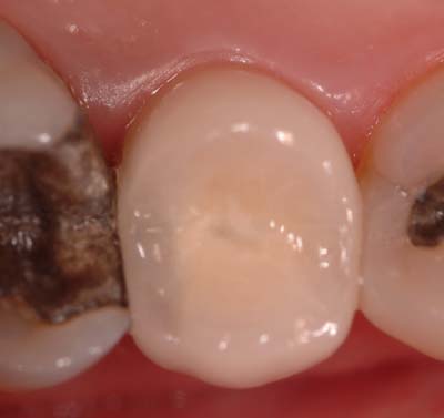
Here is the final cementation of the crown from the previously posted case. This is focused on the lingual tissue to show normalcy and healing of the laser crown lengthening procedure.

I really feel we are doing a wonderful service to these patients, and to be able to have such great results is so rewarding. Obviously, we will follow these cases, and take xrays and photos over the years to see how our results fare long term, but so far, what I have seen, is fanatastic.
Thanks everyone.
Mark
Glenn van AsSpectatorHi Bob: thanks for the post. I forgot to welcome Del to the forum.
WELCOME DEL TO THE FORUM AND IT IS GREAT TO HAVE YOUR EXPERIENCES IN ND YAG AND OTHER WAVELENGTHS HERE.
Yelling intended!!
Bob , I agree with your points and they are well taken. I thought this study was full of holes when I looked at it and it was the errors in the methods and materials section that bothered me. I immediately saw why the leakage was what it was, it was obvious but the conclusion many took out of it was that the erbium laser couldnt be used for endo.
Mark………..nice pic and nice tissue.
Do you have a picture of the prep , then we can really see the beautiful healing of the tissue .
Nicely done.
Glenn
2thlaserSpectatorGlenn, see previous post in this thread, the whole slew of pictures, including the surgery, minus the buildup are there. Thanks
Mark
Glenn van AsSpectatorHave you got the healing photos in there?
Thanks Mark
Glenn
-
AuthorPosts
