Forum Replies Created
-
AuthorPosts
-
doctorbruSpectatorHi Ron,
Went ahead and used the water spray and viola. ..first time in twenty years I had fun removing gutta percha.
Thanks for the great info.
Bruce
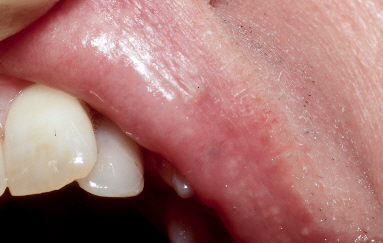
BenchwmerSpectatorHere is a four month post-op photo

Jeff
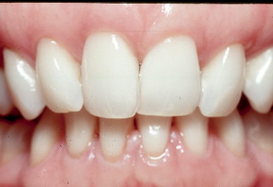
BenchwmerSpectatorPost-op after 18 months

Jeff
(Edited by Benchwmer at 6:41 pm on Dec. 14, 2005)
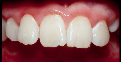
BenchwmerSpectatorStill vital and looking good going on 1 year

Jeff
jgoelzSpectatorThanks Ron, totally cool. No chace of cracking a root either. Being so much easier, I suspect we’ll be doing this in more (all) canals in the tooth & a bit deeper when we do this for core retention instead of using a post.
If one didn’t mind the sparking etc, could one do it w/out water & meerely suction the vapor? I haven’t had a chance to fiddle w GPercha-laser-interaction yet. Or was with water clearly superior operating conditions?
jgoelzSpectatorI saw an identical case, & # 18, today except there was no apico. I stuck a cone of GPercha down the fistula & it terminated at the M root apex, clearly at one of the M canals & NOT the other mesial canal ( I need another PA to use the SLOB rule to ID which mes root canal.
Since Bob & Del got my imagination running:
– if the root is not cracked
– if the canal is reasonably filled ( obviously the final 1-3 mm is not )
– if the periolase tip can be traced to the apex through the fistula
– if the coronal restoration/crown is satisfactory/ not leakingthen why not zap it? Start at the apex & continue to lase as you exit the fistula.
At what setting?
Followed by the hemostasis setting?I ask because I just can’t imagine there would be enough bacteria left in the canal in the final 1-3mm. OTOH, you say there was bacteria in the apical few mm before today to result in today’s fistula. But this case had a pre-RCT radiolucecy, but it was not draining.
I will ULTIMATELY ( not now) retreat the RCT to clean that final 1-3mm at the apex, but when I am done, that fistula (if present) is getting lased as far as it will go. It is no where near the IA nerve & I will measure the fiber to be sure.
But before I do what I stated above as the ULTIMATE/final treatment, I am going to lase only the fistula. I will report my findings. Anybody done this already? I see no harm in attempting to heal the periradicular area prior to RCS intrumentation, & who knows, maybe it’ll heal w/out re-instumenting the canals thus saving the pt that time & effort & expense. Kind of like guys doing the apico only & not re treating the canals.
Comments welcomed.
Glenn van AsSpectatorHey Bruce……..cant answer for the Cerec stuff but a few areas you can find more information about scopes are:
Global Surgical ….. http://www.globalsurgical.com
Academy of Microscope Enhanced Dentistry….http://www.microscopedentistry.com
Microscopes forum..dentaltown…….
http://www.dentaltown.com (look under magnification).Cerec has a board there as well.
Gotta run , hope that helps you a little.
Glenn van As
Glenn van AsSpectatorHey Jeff, it takes a lot to take the time to photograph the follow ups.
CLAP CLAP CLAP…..good for you.
It is neat to see.
Thanks and nice result.
Glenn
doctorbruSpectatorThanks Glenn,
What I would really like to know at this moment in time is it possible and practical to have a patient viewing a tv monitor such as the dental chair potato and still have the doctor working thru a microscope. I am having ceiling construction done in my ops and would like to plan on a microscope in the near future.
I’ll ck the sites you listed.
Thanks for posting. Looking forward to meeting you and seeing your presentaion in Feb.Bruce
BenchwmerSpectatorJohn,
Look back in this topic to 12/10/04 for a case I documented using the PerioLase.
I use LANAP settings, probe to the bone through the fistula, set the fiber to length, fire as the fiber is backed out, then one more pass at wider pulse width to set the clot.
Jeff
AnonymousSpectatorJohn, W/O water I think there would be 2 concerns-
1. Heat build up if you dilly-dallied too long.
2. Smell and sound. There can be quite a pop sound.IMO, its just is easier to add a little water than to explain to the patient what they’ll be smelling and hearing. Also it seems like you get less black discolorization (carbon) which I think would interfer w/ the bond if not removed.
beantwnSpectatorWhere do I go for the Periolase user forum??
Glenn van AsSpectatorHi Bruce…….I have a 20 inch Sony Wega in the ceiling a little behind the patient and slightly to the left.
They can then watch TV and the scope.
I know John Kois has both the Couch potato and the scope. Not sure how much he uses the scope….maybe others know.
I like to have the TV slightly off the center (my scope is ceiling mounted as well) so that the patient can still watch while I work. It works ok in my office.
My chairs are slightly off the vertical as well though.
Cya
Glenn
AnonymousSpectatorQUOTEQuote: from beantwn on 9:12 pm on Dec. 15, 2005
Where do I go for the Periolase user forum??After bootcamp you’ll recieve info from Bob,
czeqm8SpectatorAwesome result for this patient. Well done.
I think I might have a problem with this statement though.
” you need 2.6mm of biologic width. I measured 3mm from crest of the gingiva to the base of the pocket, under LA I sounded bone at 5mm from the gingival crest. That gives me 5.0mm minus 2.6mm or 2.4mm of free gingiva I can excise without violating BW.”
I think that 2.6 is an average number, and in reality patients can range from 1-4mm from the bone to the bottom of the pocket for their biological width. (correct me if I am wrong, please) I don’t think that you can use 2.6 on everyone. This patient did not rebound much, but some will (by my understanding) need more room than 2.6mm.
Am I incorrect in my thinking?
-
AuthorPosts
