Forum Replies Created
-
AuthorPosts
-
dkimmelSpectatorMark, I have been going back though some of the older post and was thinking about something you said the other day about Class 1s. I had just come back from Stewarts course and he likes using air abrasion/fissure burs to open Class 1s. Just a matter of speeding things up. What you said was something about moving quickly through Class 1s. The staff was giving me the look so I had to go and let it drop. How are you doing class one that speed things up?
David
dkimmelSpectatorAndy. The apthous ulcers were with .25 W with 11% air and no water. Just painting the surface coming from a defoused distance until I just get that whitish covering.
The herpatic lesion was .25W with 11%air and just enough water to make a mist and not droplets. I cannot remember off hand the setting for the water. Usually I do not use the water on herpatic outbreaks. In this case he had older lesions that were scabed and prevesical areas. If I used the 25W with 11% air he would get a little pin prick feeling, which scared him. With the water and coming from a defocused length in to paint he felt nothing.
I will say I was rather concerned about treating the herpatic leason because of the possible plume. Hopefully the water helped control this as well as the vaccum. Along those lines what are others doing as percations in dealing with these times of leasons?
David
SwpmnSpectatorHmmmm….Crazy stuff or SOSDD(Same Old Sh_t Different Dentist)?
 ?
? ???
???Sounds familiar to me.
Al
ASISpectatorAl,
Do I detect a distaste for the Waterlase performance?
Did you look closely at the other choices before you settled on the Waterlase?
Regards,
Andrew
SwpmnSpectatorHey Mark:
Yeah man I read in your original post where you used a T4 tip at 12% water and 9% air but what was your Power setting on your beam?
You said you used low power – one Watt, two Watts?
Al
2thlaserSpectatorRight on Al, I goofed!!! Sorry. I use .50W with a T-4, if I change to a G-6 then I use .75W.
Sorry Buddy!
Mark
Robert Gregg DDSSpectatorHey Andy,
100 usec, 3.00 watts, 20 hz, defocused, 100 joules/mm diameter lesion for your pulsed Nd:YAG.
No concern regarding plume since no vaporization is taking place. Still, high volume evacuation helps keeps the site cool. No requirement for H2O. Air is sufficient.
(Edited by Robert Gregg DDS at 9:36 am on May 2, 2003)
Robert Gregg DDSSpectatorHi Mark,
Nice case.
As another scope guy for about 5-6 years, I agree with Glenn and Allen.
But soon I’m going to post some dentin decay removed with a pulsed Nd:YAG under magnification–just for fun and so Glenn can see it IS so. 😉
Bob
Robert Gregg DDSSpectatorRon,
Kudos to you for a good literature review.
I don’t see any real surprises there:
1. relatively shallow pockets treated.
2. thermal energy killed bugsInteresting study in that J. Perio publishes studies on lasers that don’t show any real benefit to using lasers over conventional treatment.
Bob
Robert Gregg DDSSpectatorOh Yeah, Ron,
Again, good lit review.
This is a repeat of studies done years ago showing increase cell respiration using near-infrared 810-980-1064nm in a pertri dish–only using fibroblasts.
Supports the dual application of near in frared lasers to both kill pathogens and induce cell activity, proliferation and regeneration both at the time of “surgery” and through biostimulation application during the immediate post-op phase.
Bob
Glenn van AsSpectatorHey Bob………show me the money. Actually, I believe you and when you told me at the CDA that the pulsed Nd Yag wouldnt compare to the Erbium for hard tissue, that was enough for me. The NdYag is a much better laser for perio than the erbium which can also be used in a pocket but not selectively like the Nd Yag for pigmented bacteria.
Glenn
AnonymousSpectatorJust trying out a 6 paper freebie from dentalsummary.com
 and thought the rest of the visitors might find these interesting.
lagunabbSpectatorRon, thanks for posting. It sounds like a well designed double blinded study that we seldom find in the literature. Bob, are you aware of any similarly conducted experiments on Nd:YAGs for perio? I recall the Brussels (done by Nihon U) abstract that I posted but that was not double blinded. Thanks.
ASISpectatorSorry, Al. I didn’t intend to put it in a way to be critical. I share your sentiment when a piece of dental technology is not performing as it can. No matter what it is. It is a nuisance that one doesn’t need especially in a middle of a procedure.
Cheers,
Andrew
2thlaserSpectatorRemember the case on my uncle? Well here’s an interesting one on my aunt. She had chronically been complaining of a “bump” on the inside of her gum for some time, while she was at home in PA. I had her see an endodontist, who then did an apico, with no resolve. Next, he performed “flap surgery” where he thought some peiodontal pocketing was occurring, he did the procedure 2 times in a 2 month span, not finding anything, not a crack, nothing. I asked my aunt if she remembers him using magnification, either loupes (showed her mine), or a microscope, and she couldn’t remember…anyhow, here is the preop radiographs…
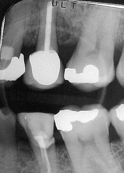
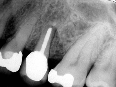
Sorry, no preop photo’s, I was VERY short on time, UNTIL I removed the crown…and here is what we saw IMMEDIATELY after removing the crown with a great white bur…..
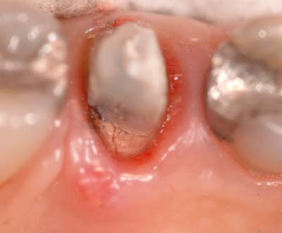
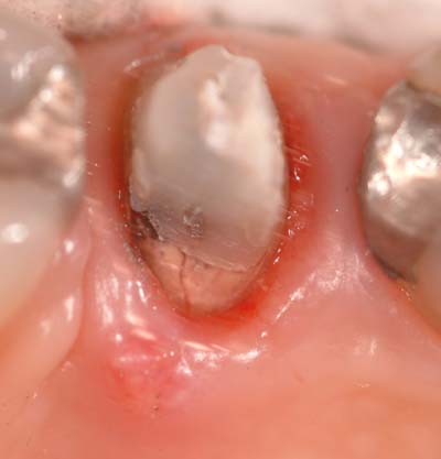
I took slightly different angles to show the fracture from the post. It was a parapost, with a composite build up. The tooth had normal occlusion, no atraumatic forces..just a chronic fracture of the root. I really think that with magnification, a surgeon would have saw the fracture upon flapping…anyone disagree? Here is the tooth extracted, note the length of the fracture, and where the bone had been destroyed by the bacteria.
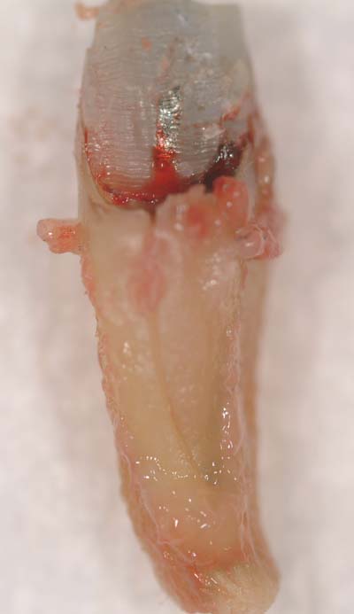
I hope you guys don’t mind me posting this. I thought it was interesting. Glenn, THIS is a good reason for magnification, don’t you think? I think a scope here would have been very beneficial.
Today, we prepped her for a bridge, she didn’t want to wait for implants…we move on!
Thanks,
Mark -
AuthorPosts
